
Fig. 5.
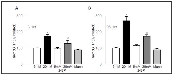
2-BP inhibits glucose-induced Rac1 activation in endothelial cells. Rac1 activation was quantified by G-LISA in the retinal endothelial cells incubated in 5mM or 20mM glucose for 3 hours (Panel A) or 96 hours (Panel B), in the absence or presence of 2-BP (100 μM). Data are expressed as percent control, and are mean ± SD from four independent experiments. 5mM and 20mM=cells incubated in 5mM glucose or 20mM glucose, respectively and Mann= cells incubated in mannitol [as an osmotic control]. *P < 0.001 vs. 5mM glucose and **P < 0.001 vs. 20mM glucose.