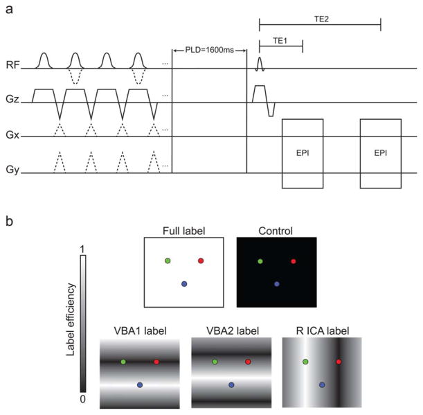
FIG. 1.
Dual echo VE-ASL and BOLD pulse sequence. a: The first half of the diagram, before the post-labeling delay (PLD), depicts the labeling pulse train for each of the pCASL labeling scenarios and the control. A series of Hanning-windowed pulses (pulse duration = 0.5 ms) were used for blood water labeling. The control scan is achieved through use of consecutive radiofrequency pulses with alternating polarity, the full label only requires the slice-select gradient, while each VE label is achieved by application of interpulse transverse (Gx and Gy) gradients. In this protocol, four separate labeling conditions were performed along with one control condition. Each pulse train is followed by a 1600 ms PLD. Acquisition occurs with two single-shot gradient echo planar imaging readouts, with TE 1 corresponding to the readout with low BOLD sensitivity (TE1 = 10.5 ms) and TE 2 corresponding to the BOLD-weighted readout (TE2 = 35 ms). b: Ideal labeling efficiency of the different ASL labeling conditions. From left to right, the top row depicts the full label (white background) and control (black background) pulses, whereas the bottom row depicts the VBA and ICA conditions: VBA1, VBA2, and R ICA.

