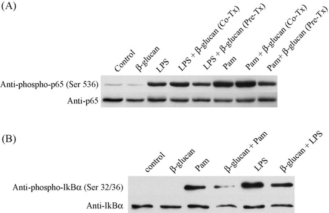
Fig. 4.
Particulate β-glucan suppresses activation of NF-κB by TLR ligands. Primary microglia (A and B) were left untreated (control) or were stimulated with LPS (1 µg/ml) or Pam3Csk4 (Pam; 1 µg/ml) for 15 min (A) or 5 min (B). Subset of cells was stimulated with β-glucan alone for 2 h (A and B) or co-stimulated with β-glucan + LPS or β-glucan + Pam3Csk4 for 15 min (A). In some experiments cells were pre-treated with β-glucan for 2 h followed by stimulation with LPS or Pam3Csk4 for 15 min (A) or 5 min (B). Cells were lysed and whole cell lysates were used to determine phospho-Ser 536 levels of NF-κB (p65) (A) or phospho-Ser 32/36 levels of IκBα (B). Equal levels of p65 and IκBα were then confirmed by stripping and reprobing the membrane using anit-p65 antibodies and IκBα antibodies respectively.

