



Summary
Toxoplasma gondii is a protozoan pathogen in the phylum Apicomplexa that resides within an intracellular parasitophorous vacuole (PV) that is selectively permeable to small molecules through unidentified mechanisms. We have identified GRA17 as a Toxoplasma-secreted protein that localizes to the parasitophorous vacuole membrane (PVM) and mediates passive transport of small molecules across the PVM. GRA17 is related to the putative Plasmodium translocon protein EXP2 and conserved across PV-residing Apicomplexa. The PVs of GRA17-deficient parasites have aberrant morphology, reduced permeability to small molecules, and structural instability. GRA17-deficient parasites proliferate slowly and are avirulent in mice. These GRA17-deficient phenotypes are rescued by complementation with Plasmodium EXP2. GRA17 functions synergistically with a related protein, GRA23. Exogenous expression of GRA17 or GRA23 alters the membrane conductance properties of Xenopus oocytes in a manner consistent with a large non-selective pore. Thus, GRA17 and GRA23 provide a molecular basis for PVM permeability and nutrient access.
Graphical abstract

Introduction
Protozoan parasites in the phylum Apicomplexa present a global human and veterinary health problem. Toxoplasma gondii, which causes toxoplasmosis, can infect a wide range of warm-blooded animals including humans and is a particular challenge to immune-compromised individuals such as AIDS patients and fetuses of infected mothers. Apicomplexan parasites spend the majority of their life cycle within host cells and have evolved diverse strategies to survive in this niche. Many, like Toxoplasma and Plasmodium, reside within a non-fusogenic parasitophorous vacuole (PV). The PV membrane (PVM) is the physical interface between PV-residing parasites and their host cells during intracellular growth, remaining intact until parasite egress. The PV protects parasites from being sensed by the host cell but it is also a potential barrier to the scavenging of nutrients from the host and to parasite effectors accessing the host. Apicomplexan parasites depend on host cells for many essential nutrients. For instance, Toxoplasma is auxotrophic for small molecules including amino acids like tryptophan and arginine, purines, and sterols [reviewed in (Coppens, 2014)]. Some, like sterols, are actively scavenged from the host cell to the PV through sterol transporters on the PVM (Ehrenman et al., 2010). Transporters for small metabolites have been identified on the Toxoplasma plasma membrane but not on the PVM (Landfear, 2011). The Toxoplasma, Plasmodium, and Eimeria PVMs are selectively permeable, allowing passive, charge-independent diffusion of small molecules up to 1300-1900 Da into the PV, presumably through pore-like molecules on the PVM (Desai and Rosenberg, 1997; Schwab et al., 1994; Werner-Meier and Entzeroth, 1997). This is the putative mechanism by which the parasite within the PV gains access to host metabolites or effluxes toxic metabolic byproducts like lactate into the host cell. The molecular identity of the pores that grant this PVM permeability is unknown.
Toxoplasma partially circumvents the PVM barrier by secreting proteins into the host cell before and during invasion before formation of the PV from secretory organelles that include rhoptries and dense granules. Secreted effectors play a critically important role in Toxoplasma pathogenesis, modulating host signaling and creating a hospitable environment in the PV [reviewed in (Hunter and Sibley, 2012; Kemp et al., 2012)]. Unlike the invasion-secreted rhoptry proteins (ROPs), dense granule proteins (GRAs) are also secreted post-invasion (Karsten et al., 1998).
Plasmodium exports proteins beyond its PVM into the erythrocyte cytosol. EXP2 is the putative protein-conducting pore of the PTEX (Plasmodium translocon for exported proteins) complex that mediates the essential export of secreted proteins containing a PEXEL (Plasmodium export element) sequence motif (de Koning-Ward et al., 2009). There are also PEXEL-independent mechanisms of protein export (Gruring et al., 2012; Siau et al., 2014) that utilize the same PTEX machinery (Beck et al., 2014; Elsworth et al., 2014). In Toxoplasma, post-invasion protein export into the host cell has only been reported for GRA16 and GRA24 (Bougdour et al., 2013; Braun et al., 2013). Most characterized Toxoplasma GRA proteins are involved in organizing the structure and environment of the PV or associated with the PVM but others such as GRA15, GRA16, and GRA24 play a role in modulating host cell signaling, suggesting that there may be more GRA proteins trafficked beyond the PVM after invasion (Bougdour et al., 2013; Mercier et al., 2005; Rosowski et al., 2011).
We sought to investigate the molecular interaction between the Toxoplasma PV and the host cell by studying a PVM-localized GRA protein with homology to Plasmodium EXP2.
Results
GRA17 is a secreted GRA protein required for normal PV morphology
Since EXP2 has been implicated in forming a molecular-translocation channel in the P. falciparum PVM, we searched for homologs in the Toxoplasma genome, and found two genes encoding proteins with predicted signal peptides: the uncharacterized TGGT1_222170 and TgGRA23 (TGGT1_297880), an identified GRA gene of unknown function (Masatani et al., 2013). TGGT1_222170 and TgGRA23 share 30% amino acid sequence similarity with each other and 26% and 22% similarity with PfEXP2, respectively (Fig. S1A). Homologs of these proteins, including Sarcocystis muris DG32 (Freyer et al., 1999), were identified only in Apicomplexa that replicate within a PV (Table 1). Except for Plasmodium spp. EXP2, none of these genes have been functionally characterized but due to its similarity to GRA23, we provisionally named TGGT1_222170, “GRA17”. There is only one gene similar to TgGRA17 or TgGRA23 found in Eimeria spp. and Plasmodium spp. but we found two distinct homologs in the genomes of other sequenced PV-residing Apicomplexa that are grouped in the family Sarcocystidae (Table 1; Fig. S1B). We found that GRA17 and GRA23 are conserved proteins with high gene transcription levels across a global range of Toxoplasma strains (Melo et al., 2013; Minot et al., 2012).
Table 1. GRA17-like genes are unique to PV-residing Apicomplexa.
The peptide sequences of GRA17 and GRA23 were used as bait in searches against Apicomplexan organisms in EuPathDB.org using the BLASTP 2.0 algorithm or TBLASTN 2.2.26. Subsequent hits receiving a P value less than 0.005 were identified as similar to GRA17 and themselves used as baits in subsequent searches.
| Organism | GRA17-like genes | Gene names | PV? |
|---|---|---|---|
| Toxoplasma gondii | 2 | GRA17 (TGGT1_222170), GRA23 (TGGT1_297880) | yes |
| Neospora caninum | 2 | NCLIV_005560, NCLIV_006780 | yes |
| Hammondia hammondi | 2 | HHA_222170, HHA_297880 | yes |
| Sarcocystis spp. | 2 | S. neurona Aa, S. neurona B; S. muris DG32b | yes |
| Eimeria spp. | 1 | E. acervulina EAH_00026470 (DG32) and E. mitis EMH_0012530 (DG32)c | yes |
| Plasmodium spp. | 1 | P. falciparum PF3D7_1471100 (EXP2), P. knowlesi PKH_123420 (EXP2), P. reichenowi PRCDC_1470300 (EXP2) | yes |
| Babesia bovis | 0 | NA | no |
| Cryptosporidium spp. | 0 | NA | no |
| Theileria spp. | 0 | NA | no |
| Gregarina niphandrodes | 0 | NA | no |
The S. neurona genome is sequenced but unannotated, so the two GRA17-like genes were arbitrarily named “A” and “B”.
The S. muris genome has not been sequenced so we cannot determine whether there is a second GRA17-like gene.
GRA17-like genes were identified for all Eimeria and Plasmodium species whose genomes have been sequenced but, for brevity, only a selection of these were included in this table.
Besides strongly predicted α-helices, GRA17 and GRA23 lack sequence motifs that suggest a functional role. To characterize them, we individually disrupted the GRA17 and GRA23 genes and we complemented the ΔGRA17 strain with either HA-tagged GRA17 or Toxoplasma-codon optimized P. falciparum EXP2 transgenes that we will refer to as “ΔGRA17+GRA17-HA” and “ΔGRA17+PfEXP2-HA” (Figs. S2A-E). The ΔGRA17 strain was also complemented with GRA23-HA-FLAG expressed from either the putative gra23 promoter, “ΔGRA17+GRA23-HFlow” or from the strong gra1 promoter “ΔGRA17+GRA23-HFhigh”; the differing expression levels in these strains were verified by Western blot (Fig. S2F). We infected human foreskin fibroblasts (HFFs) with tachyzoites (the fast-replicating stage of Toxoplasma), fixed and analyzed the sample by deconvolution immunofluorescence (IF) microscopy. The ΔGRA17+GRA17-HA strain was used for many co-staining experiments as it does not express GFP, and the GRA17-HA localization was invariant across all the strains (data not shown). We observed that GRA17-HA is intra-vacuolar, associated with the PVM and possibly the tubulo-vesicular network (TVN) based on its partial co-localization with GRA7 (a PVM and TVN marker) and GRA4 (a TVN marker) especially around the periphery of the PV (Fig. 1A). GRA23 has previously been characterized as a PVM-localized GRA (Masatani et al., 2013) and to observe its relative co-localization with GRA17 we generated a strain that co-expresses GRA17-V5 and GRA23-HF (the tachyzoites express GFP) and observed substantial co-localization around the PVM (Fig. 1A, bottom panel; Fig. S2G), however the localization of GRA17-HA appeared localized to discrete foci relative to the more continuous pattern of GRA23-HF around the PVM. PfEXP2-HA localized to the PVM in the ΔGRA17 strain and the localization of GRA23-HF was unaffected by the absence of GRA17 in the ΔGRA17+GRA23-HFlow strain (Fig. S2H). We confirmed that GRA17 is a GRA by observing partial co-localization of GRA17 and GRA7 in extracellular tachyzoites (Fig. S2I). We raised a specific antibody against GRA17 that detected a 19 kDa band that is absent in the ΔGRA17 strain (Fig. S3A, lanes 1-4). We tested GRA17 association with the PVM by fractionating HFFs infected with WT+GRA17-HA by ultracentrifugation and extracting a membrane-enriched fraction with either PBS or sodium carbonate. Sodium carbonate treatment extracts peripherally attached proteins such as GRA1 but more tightly membrane-associated proteins like GRA7 remain in the insoluble fraction (Neudeck et al., 2002; Sibley et al., 1995). We found that in contrast to GRA1, GRA17 was only minimally extracted by sodium carbonate and that the majority remained in the insoluble fraction (Fig. 1B). Thus, the majority of GRA17 is tightly associated with the PVM. A similar fractionation pattern for GRA23 has been reported (Masatani et al., 2013).
Figure 1. GRA17 associates with the PVM and affects PV morphology.

(A) Human foreskin fibroblasts (HFFs) were infected with the indicated Toxoplasma tachyzoite strains for 24 h, fixed and subjected to IF with the antibodies indicated. The WT+GRA17-V5/GRA23-HA-FLAG also expresses cytosolic GFP. The fluorescent images represent a single deconvoluted focal slice. Brightfield images were taken with differential interference contrast (DIC) microscopy (scale bar = 10 μm). (B) HFFs were infected with WT+GRA17-HA tachyzoites for 24 h, mechanically lysed and a membrane-enriched fraction was treated with either PBS or sodium carbonate and centrifuged to obtain pelleted (P) and soluble (S) fractions and subjected to SDS-PAGE. The fractions were Western blotted as indicated. (C) HFFs were infected with the indicated strains for 36 h and imaged live by phase-contrast microscopy (scale bar = 25 μm).
We observed that a significant portion of PVs in the ΔGRA17 strain were grossly enlarged relative to the number of parasites in the PV, whereas the ΔGRA23 PVs are similar to WT (Fig. 1C). Furthermore, the ΔGRA17-complemented strains, ΔGRA17+GRA17-HA and ΔGRA17+PfEXP2-HA, completely rescued this aberrant morphology. We were unable to generate a ΔGRA17/ΔGRA23 strain but we were able to delete the GRA23 gene from the ΔGRA17+GRA17-HA strain, which has low GRA17 expression (Fig. S3A, lanes 2, 8). We observed that this ΔGRA17/ΔGRA23+GRA17-HA strain mimics the aberrant PV morphology of the ΔGRA17 strain (Fig. 1C), providing evidence of functional overlap between GRA17 and GRA23. Furthermore, the ΔGRA17 strain complemented with high (but not lower) expression of GRA23-HF partially suppressed this phenotype, with infrequent enlarged vacuoles observed (Fig. 1C). We examined whether there were other genes involved in a compensatory role with the ΔGRA17 strain by sequencing the total mRNA from bone marrow-derived mouse macrophages 4 hours post-infection with WT, ΔGRA17, ΔGRA23, and ΔGRA17+GRA17-HA strains and mapping the sequences to the Toxoplasma genome. As previously observed, GRA17 expression in the ΔGRA17+GRA17-HA strain was 2.5-fold less compared to WT despite its ability to functionally rescue the ΔGRA17 strain (Fig. S3B). GRA23 expression levels in the ΔGRA17 strain were 1.8-fold increased over wild-type levels and GRA17 levels were not increased in the ΔGRA17 strain (Fig. S3B). We found five additional, uncharacterized putative signal peptide-containing proteins that were uniquely up-regulated in the ΔGRA17 strain (Table S2).
GRA17 is involved in the movement of small molecules between the host and PV
Since PfEXP2 can rescue a ΔGRA17 phenotype, we wondered whether GRA17 or GRA23 plays a role in protein export beyond the PVM. The GRA proteins GRA16 and GRA24 localize to the host nucleus with increasing intensity over the time of infection, peaking at 20 hours, indicating that they are exported across the PVM after the PV has been formed (Bougdour et al., 2013; Braun et al., 2013). We tagged the endogenous copy of GRA24 with HA-FLAG (HF) in the WT, ΔGRA17, and ΔGRA23 strains. We also transiently transformed GRA16-HF into WT, ΔGRA17, and ΔGRA23 extracellular tachyzoites with which we immediately infected HFFs. We then fixed the infected cells at early and late time points post-infection. Across all strains we observed a temporal increase from low to high levels of GRA16-HF and GRA24-HF staining in the host nucleus (Figs. 2A, S3C). Thus, neither GRA17 nor GRA23 are individually required for GRA16 and GRA24 export beyond the PVM.
Figure 2. GRA17 is required for the transfer of small molecules into the PV.

(A) Indicated tachyzoite strains endogenously tagged with GRA24-HA-FLAG were used to infect HFFs and the samples were fixed at the indicated time-points, stained with Hoechst dye (red), anti-HA (green), and overlaid onto a DIC image. For each channel, the exposure time is the same. This experiment was performed twice. (B) Sub-confluent HFFs were infected with the indicated strains for 24 h after which they were pulse labeled with CDCFDA (green) for 10 min before the cells were imaged live. White arrows indicate intra-vacuolar fluorescence and the red arrow indicates the absence of intra-vacuolar fluorescence (left panel; scale bars = 10 μm). The percentage of CDCF-fluorescent vacuoles was quantified for each strain and averaged across experiments (right panel). A minimum of 50 vacuoles per strain was counted for each replicate. Error bars represent ±SD and the numbers above the bars of the graph indicate the number of experimental replicates for each condition. Unpaired two-tailed t tests compared to WT (*** P=3×10⁻⁷; ** P=0.002) or ΔGRA17 (### P≤0.00001, ##P=0.01) were performed. (C) Confluent HFFs were infected with the indicated strains for 24 h after which either Lucifer yellow or Dextran3000-Alexa 488 were scrape-loaded into the cells and imaged live immediately. Intra-PV fluorescence was scored relative to the fluorescence of the individual host cell as present, intermediate, or absent as a percentage of total vacuoles (Fig. S3D) and averaged across 3 experimental replicates (Error bars = ±SD).
While GRA17 and GRA23 do not appear to affect protein transport across the PVM, we asked if they function as a pore for small molecules and metabolites. We used the properties of the vital dye 5-(and-6)-Carboxy-2′,7′-Dichlorofluorescein Diacetate (CDCFDA) to test this hypothesis. CDCFDA is membrane permeable and non-fluorescent until it enters living cells, where intracellular esterases convert it into the fluorescent, membrane-impermeable 5-(and-6)-Carboxy-2′,7′-Dichlorofluorescein (CDCF). The molecular weight of the CDCF fluorophore (445.2 Da) is less than the established size exclusion limit of the Toxoplasma PVM (Schwab et al., 1994), so we predicted that it should passively enter into the PV through the PVM but be unable to permeate through the tachyzoite plasma membrane (Schwab et al., 1994). We infected HFFs with the WT, ΔGRA17, ΔGRA17+GRA17-HA, ΔGRA23, ΔGRA17+PfEXP2-HA, ΔGRA17/ΔGRA23+GRA17-HA, and ΔGRA17+GRA23-HFlow/high) strains for 24 hours and then added a pulse of CDCFDA to the cell culture medium before washing away the dye and subsequently imaging the living cells. As predicted, we observed CDCF-fluorescence intensity equivalent to the host cytosol within nearly all the PVs of the WT, ΔGRA17+GRA17-HA, ΔGRA17+PfEXP2-HA, and ΔGRA23 strains but excluded from the tachyzoites themselves (Fig. 2B, white arrows). By contrast, 36% of the PVs in the ΔGRA17 strain had equivalent intra-PV CDCF-fluorescence compared to the host cytosol (P=4×10⁻⁸ to WT; Fig. 2B, red arrow and right panel), suggesting a role for GRA17 involving the passage of small molecules into the PV. We observed a similar phenotype for the ΔGRA17/ΔGRA23+GRA17-HA strain, with CDCF-fluorescence in 51% of PVs (P=0.002 to WT and no statistical difference to ΔGRA17; Fig. 2B, right panel). In contrast to the ΔGRA17+GRA23-HFlow strain, which was similar to the ΔGRA17 strain (45% intra-PV CDCF-fluorescence; P=0.002 from WT) we observed a partial rescue of dye transfer with the ΔGRA17+GRA23-HFhigh strain with CDCF-fluorescence in 73% of PVs that was statistically distinct from the ΔGRA17 strain (P=0.01; Fig. 2B), further suggesting a synergistic role for GRA23 in small molecule permeability. Since we observed PVs of varying permeability within the same host cell (Fig. 2B, ΔGRA17), the GRA17-mediated effect on PVM permeability to the small molecule fluorophore is PV-autonomous and independent of host intracellular fluorophore concentration.
We confirmed this phenotype by introducing the membrane-impermeable dyes Lucifer Yellow (522 Da) or Dextran (3000 Da)-Alexa-488 into infected cells by the technique of scrape-loading (McNeil et al., 1984). Since cellular dye uptake varies around the scrape, there was a large degree of heterogeneity of both intracellular and intra-PV dye intensities. We scored the intra-PV dye intensity relative to its host cell dye intensity as “present”, “absent”, or “intermediate” (Fig. S3D). We found that the scrape-loaded Lucifer Yellow results were similar to the CDCFDA results: in contrast to >90% of WT, ΔGRA17+GRA17-HA, and ΔGRA23 PVs, only 22% of ΔGRA17 PVs were scored as having fluorescence fully equivalent to their host cells (Fig. 2C). When the larger Dextran (3000 Da) dye was scrape-loaded into infected cells, we found that most of the PVs of both WT and ΔGRA17 failed to uptake the dye, consistent with the established Toxoplasma PVM size-exclusion limit (Schwab et al., 1994). These results provide further evidence that PfEXP2 can rescue ΔGRA17 phenotypes and for functional overlap between GRA17 and GRA23.
The absence of GRA17 results in highly dynamic, unstable PV
We found that fixatives altered the appearance and numbers of the enlarged, aberrant ΔGRA17 PVs (data not shown) so we imaged the ΔGRA17 strain live over an extended time period post-infection to observe the dynamics of these PVs. Nearly all of the PVs we observed by live imaging appeared enlarged at some point during the experimental time course. We also observed many enlarged PVs “collapsing”, where the tachyzoites within them turn opaque and cease to divide. As an example, we present a multiple infected host cell where all but one PV collapses over a 5:30 hour time course (Fig. 3A; Video S1). The remaining intact PV within this cell remained enlarged and continued to divide, ultimately egressing from the cell at 62:00 hours (Video S2). We have also observed PV collapse in singly infected cells (Fig. S4A). Furthermore, we observed dynamic alteration of the size and shape of the vacuoles as exemplified by a PV that becomes grossly enlarged to the size of the entire host cell before ultimately egressing (Fig. S4B; Videos S3, S4). Since the tachyzoites within these enlarged PVs divide normally and we regularly observe egress, it indicates that they are viable.
Figure 3. GRA17 affects the stability of Toxoplasma PVs.

(A) Sub-confluent HFFs on were infected with ΔGRA17 tachyzoites and imaged live. Images obtained by phase-contrast microscopy are presented at 30 min time points from 19:30 h through 25:00 h post-infection. Matching colored arrowheads indicate the collapse of indicated PVs in consecutive time points (scale bar = 10 μm). (B) Confluent monolayers of HFFs were infected with 100 tachyzoites of each indicated strain for 4 days. For every experiment, the area of at least 30 plaques from each strain was measured (Fig. S5A) and the mean area was normalized to WT. Each bar of the graph represents an average of means across experimental replicates (n=8 for all conditions except for ΔGRA17+GRA17-HA, where n = 3). Error bars represent ±SD and paired two-tailed t tests were performed comparing each condition to WT to assess statistical significance (***; P<0.0001).
In concordance with the PV collapse, we observed that ΔGRA17 tachyzoites grew more slowly than WT tachyzoites. We measured this effect by quantifying the mean area of plaques formed in HFF monolayers by infection with the ΔGRA17 and ΔGRA23 strains relative to WT. These assays measure the overall growth of the parasites in vitro. We found that ΔGRA17 formed plaques that were 53% the size of the WT strain (P<0.0001; Figs. 3B, S5A) and the ΔGRA17+GRA17-HA strain fully rescued this growth defect. The ΔGRA23 strain formed plaques that were slightly smaller than the WT strain but this effect was not significant (P=0.21; Figs. 3B, S5A). In contrast to the smaller plaques made by the ΔGRA17 strain, we observed that a strain that overexpresses GRA17-HA (WT+GRA17-HA; Fig. S3A, lanes 9, 10) formed plaques that were 67% larger than WT (P<0.0001; Figs. 3B, S5A), implying that excess GRA17 can accelerate growth of the parasite. The collapsed PVs with non-dividing tachyzoites suggest a basis for the slow growth of the ΔGRA17 strain in vitro.
GRA17 and GRA23 affect the in vivo growth and virulence of Toxoplasma
We examined whether the effects of GRA17 on overall Toxoplasma proliferation in vitro would manifest in vivo. We infected CD-1 outbred mice intraperitoneally (i.p.) with 100 tachyzoites of the WT, ΔGRA17, ΔGRA17+GRA17-HA, and ΔGRA23 strains. We also infected an additional cohort with 1000 tachyzoites of the ΔGRA17 strain. None of the mice infected with the WT, ΔGRA23 and ΔGRA17+GRA17-HA strains survived beyond 12 days post-infection (Fig. 4A), consistent with the reported hyper-virulence of the type I genetic background of our strains (LD100 = ∼1 tachyzoite). By contrast, all of the mice infected with either the low or high dose of the ΔGRA17 tachyzoites survived. All of these mice were seropositive and were resistant to subsequent challenge with 10,000 WT (type I) tachyzoites (Fig. 4A, arrow). Subsequently, we infected cohorts of CD-1 mice with 100 tachyzoites each of the ΔGRA17, ΔGRA17+GRA17-HA, ΔGRA17+PfEXP2-HA, and ΔGRA17/ΔGRA23+GRA17-HA strains and monitored both body weight and survival. We found that none of the mice infected with ΔGRA17+GRA17-HA or ΔGRA17+PfEXP2-HA survived past 12 days post-infection, whereas all of the mice infected with the ΔGRA17 or ΔGRA17/ΔGRA23+GRA17-HA strains survived the duration of the experiment (Fig. 4B, top). In contrast to the mice infected with ΔGRA17 parasites, those infected with ΔGRA17/ΔGRA23+GRA17-HA experienced a significant drop in body weight and deterioration in body condition (e.g. hunched stance and ruffled fur) during the course of infection before recovering fully by 30 days post-infection (Fig. 4B, bottom). Thus PfEXP2 can rescue the loss of GRA17 in vivo and the deletion of GRA23 with low GRA17 expression renders Toxoplasma avirulent. Since the type I strain is hyper-virulent in mice, we tested the accelerated growth in vivo by overexpressing GRA17 in the far less virulent type III strain (LD50=105 tachyzoites), which also accelerated its in vitro growth (Fig. S5B). We infected BALB/c mice with this strain and a WT (type III) strain. We found that all the infected mice survived but the mice infected with type III+GRA17-HA had a higher parasite burden (Fig. S5C), lost more weight (Fig. S5D), a worse body condition (e.g. hunched posture and ruffled coat), and a higher brain tissue cyst count in some but not all of this cohort (Fig. S5E).
Figure 4. GRA17 affects the virulence and in vivo proliferation of Toxoplasma.

(A) Cohorts of five female CD-1 mice (aged 6-8 wks) were each infected with 100 tachyzoites (one additional cohort was infected with 1000 tachyzoites of ΔGRA17) intraperitoneally with each type I (RH) strain and survival was monitored. At day 30, the surviving ΔGRA17 mice from the 100 tachyzoites infection were re-infected with 104 WT tachyzoites and survival was monitored (arrow). (B) Infection of mice was carried out as above and survival was monitored for 34 days post-infection (top panel). The mice were weighed throughout the infection and plotted as an average of the change in body weight for each cohort, where 100% body weight is the day before infection (bottom panel). Error bars indicate +SD and single asterisks indicate P<0.05 statistical significance assessed at that time point compared to ΔGRA17 by a student's t test.
GRA17 and GRA23 are synergistically required for growth
We were able to detect ΔGRA23 phenotypes only in the background of significantly reduced GRA17 expression and the overexpression of GRA23-HF in the ΔGRA17 background was able to partially rescue ΔGRA17 phenotypes, suggesting GRA17 and GRA23 operate synergistically. While we were able to generate multiple, independent clonal tachyzoite isolates of ΔGRA17/ΔGRA23+GRA17-HA, we could not generate a ΔGRA17/ΔGRA23 strain in parallel by multiple methods. In order to determine whether a ΔGRA17/ΔGRA23 strain is viable, we used the CRISPR gene disruption technology developed for Toxoplasma (Shen et al., 2014; Sidik et al., 2014). We chose target strains where GRA17-HA is the only species of GRA17 present so we could follow its fate by IF microscopy. As a test, we co-transformed the ΔGRA17/ΔGRA23+GRA17-HA strain with Cas9-3×FLAG, a guide RNA (gRNA) to target protospacer sequences early within the GRA17 coding sequence as well as a double-stranded oligonucleotide donor template containing a nonsense mutation near the predicted Cas9-cleavage site. We co-transformed donor templates with our gRNAs since our target strain background is ΔKu80, which eliminates non-homologous end-joining in favor of homologous recombination (Fox et al., 2009). We used three different gRNA/donor template pairings, fixed at 24 h and 48 h, and observed these gRNAs led to similar GRA17-HA loss relative to no loss with a non-specific gRNA (Fig. 5A top panel; Fig. S6).
Figure 5. GRA17 and GRA23 are synergistically required for growth.
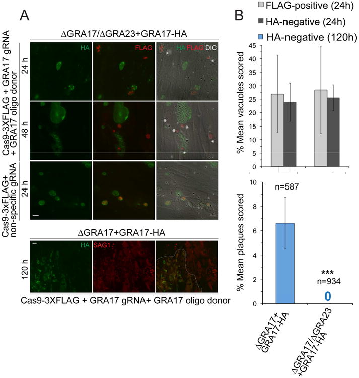
(A) Confluent HFFs were infected with ΔGRA17/ΔGRA23+GRA17-HA or ΔGRA17+GRA17-HA parasites that were transiently co-transformed with Cas9-3×FLAG and either non-specific or GRA17-specific gRNAs along with GRA17 donor oligonucleotide templates containing a nonsense mutation. The infected cells were fixed at 24 h, 48 h, or 120 h at different MOIs and subjected to IFA with anti-HA and anti-FLAG antibodies (sample image, top left panel; scale bar = 10 μm) or anti-HA and anti-SAG1 antibodies (sample image, bottom left panel; scale bar = 25 μm). White asterisks indicate vacuoles where HA-signal is absent. The dashed line distinguishes between the two plaques. (B) The percentage of Cas9-3×FLAG-positive vacuoles and GRA17-HA-positive vacuoles out of the total were scored and averaged (top). In total, 750 vacuoles each were scored across three experimental replicates. The percentage of Cas9-3×FLAG-positive plaques and GRA17-HA-positive plaques out of the total were scored and averaged (bottom panel). The number, n, over each bar graph refers to the total number of plaques scored in total for each condition across three experimental replicates. Error bars represent ±SD. Statistical significance was determined using a two-tailed Fisher's Exact Test comparing the total number of HA-negative vacuoles and plaques for both conditions (***, P<0.0001).
After transformation of both the ΔGRA17/ΔGRA23+GRA17-HA and ΔGRA17+GRA17-HA strains with Cas9-3×FLAG, a GRA17-specific gRNA/donor [(residues 29-36)/donor template (Leu32stop)] and subsequent infection of HFFs, we fixed infected cells early to compare the initial loss of GRA17-HA signal in the population (24 hours) to late, when the parasites will form plaques in the host cell monolayer (120 hours). Both target strains were transformed at an equivalent rate as measured by Cas9-3×FLAG detection (∼27-28%; Fig. 5B top panel). We also found that ΔGRA17+GRA17-HA and ΔGRA17/ΔGRA23+GRA17-HA had comparable loss of GRA17-HA signal at 24 hours post-infection (23% and 26% out of the total population, respectively; Fig. 5B top panel). By contrast, we found that 6.6% (of 584 total plaques scored) of the ΔGRA17+GRA17-HA plaques were GRA17-HA negative at 120 hours post-infection compared to 0% (of 934 plaques scored) in the ΔGRA17/ΔGRA23+GRA17-HA strain (P<0.0001; Figs. 5A bottom panel and 5B bottom panel). In parallel, we serially diluted the transformants into HFFs to isolate tachyzoite clones: 2/20 clones from the ΔGRA17+GRA17-HA strain were GRA17-HA negative compared to 0/120 clones from the ΔGRA17/ΔGRA23+GRA17-HA strain. Taken together, our results strongly indicate that GRA17 and GRA23 are synergistically required for Toxoplasma growth.
GRA17 and GRA23 Alter the Membrane Conductance of Xenopus Oocytes
We expressed GRA17 and GRA23 in Xenopus oocytes to see if they can alter membrane permeability consistent with the insertion of large, relatively non-selective pores into the cell membranes. The expression of such a non-selective pore makes several experimentally testable predictions. The first is that expression will be detrimental to cells, resulting in significant cell death due to loss of cell contents and inability to maintain volume homeostasis. When varying levels of GRA17 and GRA23 cRNAs were injected into Xenopus oocytes, a dose-dependent increase in cell lysis was observed relative to water-injected cells or to GRA15 cRNA, a GRA protein that localizes to the PVM (Rosowski et al., 2011) but which is not predicted to affect membrane permeability (Fig. 6A). HA-tagged versions of GRA17, GRA23, and GRA15 were detected in total membrane fractions isolated from cRNA-injected oocytes (Figs. S7A,B) and GRA17 and GRA23 were also observed localized to the oocyte plasma membrane in fixed histology sections (Fig. S7C). The second prediction is that increased membrane permeability through expression of putative non-selective pores would cause a depolarizing shift in membrane resting potential prior to cell lysis and GRA17 and GRA23 both demonstrated this shift relative to water- or GRA15-injected oocytes (Fig. 6B). Finally, the current to voltage relationship of the net membrane current for a large non-selective, constitutively open pore should result in an increased linear current-voltage relationship (Fig. 6C), a depolarization of the reversal potential (“Vrev”; Fig. 6D, top panel) and an increased conductance (“G”; Fig. 6D, bottom panel) for GRA17 and GRA23 relative to water- or GRA15-injected oocytes, which we observed. The results of these exogenous expression studies are strongly consistent with the hypothesis that GRA17 and GRA23 can form non-selective high conductance pores when exogenously expressed in cell membranes. We injected a separate batch of Xenopus oocytes with PfEXP2-HA cRNA and as with the Toxoplasma GRAs found that it was present in total membrane and cytosolic fractions (Fig. S7D). Relative to GRA17 and GRA23, PfEXP2 cRNA-injected oocytes had a more modest decrease in survival (73% compared to 93% of water-injected oocytes; Fig. S7E). There was no statistically significant difference between PfEXP2 cRNA-injected oocytes and water-injected control resting membrane potential (Fig. S7F), however the control oocytes had a significantly depolarized resting membrane potential compared to the previous batches of oocytes used for the Toxoplasma GRAs (Fig. 6B), making direct comparison difficult. We did observe a statistically significant increased conductance (G) change in PfEXP2 cRNA-injected oocytes (Figs. S7G and H), which is consistent with PfEXP2 forming a large membrane pore. Further work is needed to characterize the role of PfEXP2 as a membrane pore.
Figure 6. GRA17 or GRA23 alter the membrane conductance properties of Xenopus oocytes.
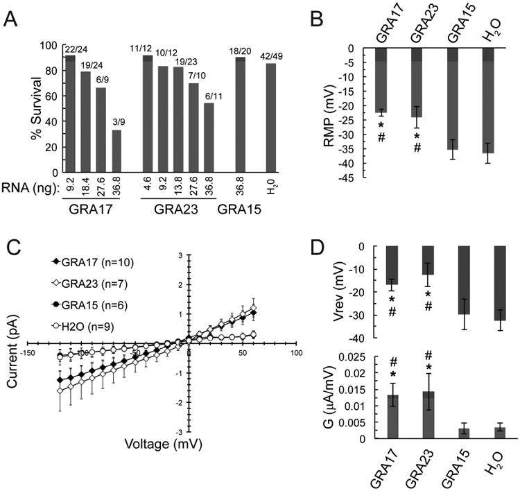
(A) Percentage survival of Xenopus laevis oocytes injected with indicated amounts of mRNA or H2O in a dose-dependent manner 3 days post-injection. Survival was based on a morphological appearance of the oocytes. Fractions on top of each bar graph represent the number of oocytes that survived out of the total injected. (B) Average resting membrane potentials (RMP) of surviving oocytes. (C) Average membrane currents measured from a holding potential of -90 mV potentials of -120 mV to +50 mV were applied for 2 seconds. Peak currents from each applied voltage were used to generate an I-V relationship. (D) Average conductance (G, bottom) and the reversal potential (Vrev, top) were computed by fitting the I-V relationship as a straight line. For all above sub-figures, data are shown as the mean ±SE. Confidence levels were calculated using Student's paired t-test, where * signifies P<0.05 when compared to H2O-injected oocytes and # signifies P<0.05 when compared to GRA15-injected oocytes.
Discussion
Intracellular pathogens that reside within vacuoles face a challenge of scavenging nutrients and modulating the host cell beyond their vacuole membrane. The existence of putative pores providing selective permeability to small molecules in the Toxoplasma and Plasmodium PVMs had been described (Desai and Rosenberg, 1997; Schwab et al., 1994) and now we have provided a molecular basis for this phenomenon in any Apicomplexa. Small molecule flow between the host cytosol and PV plays a critical role in tachyzoite growth and survival and its reduction may result in either restriction to nutrients or the inability of toxic metabolic byproducts to efflux out of the PV. Overexpressing GRA17 leads to more rapid growth, suggesting that nutrients scavenged by Toxoplasma via GRA17 may be limiting factors for normal growth of the parasite. An important question is how the ΔGRA17 strain remains viable in vitro when it is starved from access to small molecules. A hint as to why some parasites retain viability lies in the aberrant morphology of the ΔGRA17 PVs. The tachyzoites within the enlarged vacuoles are viable as they replicate and egress. The aberrant PVs may then be the byproduct or consequence of a compensatory mechanism for GRA17 absence. Compensation may include a change in the osmolarity or other structural forces, resulting in PV enlargement. The collapsed PVs may be due to ineffective compensation to reduced access to small molecules or to overcompensation that leads to PV damage. PV collapse, as well as the sensitivity of the enlarged PVs to fixatives, hints at the dynamism and fragility of the ΔGRA17 PVs and implies a precarious balance of compensatory forces.
A prime candidate for a compensatory molecule is GRA23 based on the body of evidence we have presented. One model is that GRA17 and GRA23 may form a variety of homo- or hetero-multimers whose biophysical properties may differ depending on the composition of the complex. Alternatively, GRA17 and GRA23 may function on the PVM separately, as monomers or otherwise, having partially overlapping but ultimately separable functions. GRA17 and GRA23 may function similarly to α-helical pore-forming molecules like colcins or the pro-apoptotic protein Bax that can exist in either a water-soluble state or as an integral membrane pore that alters permeability [reviewed in (Parker and Feil, 2005)]. Particularly intriguing are Bax-derived small α-helical peptides that can homo-multimerize within the membrane via a toroidal mechanism that does not require direct protein-protein interactions but instead contains proteins interspersed with lipids (García-Sáez et al., 2006; Qian et al., 2008). We have immunoprecipitated both GRA17 and GRA23 from PVM-containing fractions in the cell (data not shown) but we have so far not been able to identify specific interacting proteins. Our mRNA sequencing experiment did identify several other genes encoding putatively secreted protein products that are uniquely up-regulated in the ΔGRA17 strain but none of these have been functionally characterized to date. Thus, future work is required to define the exact molecular functions of GRA17 and GRA23 as well as identify associated molecular factors.
The observation that Plasmodium EXP2 can rescue ΔGRA17 phenotypes hints at a possible role of GRA17 or GRA23 in protein export in Toxoplasma. However, GRA16 and GRA24 accumulation in the host nucleus via post-invasion in both the ΔGRA17 and ΔGRA23 strains argues against such a role, although GRA17 and GRA23 may have redundant functions in protein export. It will be difficult to distinguish between a potential defect in export in the absence of both proteins versus parasites that are dead or dying and thus unable to actively export protein. Ultimately, very little is known about Toxoplasma protein export and there are currently only two reported exported proteins to test as candidates. Even though Plasmodium EXP2 has not been formally shown to be the translocation pore for protein export, it begs the question of whether it serves a dual role of nutrient pore and protein channel since EXP2 is the only GRA17-like protein in Plasmodium spp. The aforementioned α-helical pore-forming proteins can form pores big enough for peptides in many cases (Parker and Feil, 2005), suggesting this dual functionality is feasible. It is possible that the other proteins in the PTEX complex may direct the function of EXP2 from a potential nutrient pore towards a role in protein export. GRA17 and GRA23 may help elucidate another longstanding question of Toxoplasma biology concerning the sporozoite, the sexually derived stage found in the oocyst. After excystation from the oocyst and invasion of the host cell, the sporozoite forms a unique vacuole (PV1) in which the sporozoite undergoes no replication and ultimately egresses from the PV1 to form a second vacuole (PV2) in the same cell where it completes its conversion to the tachyzoite stage and undergoes replication. Unlike tachyzoite PVs and PV2, the PV1 is impermeable to small molecules and devoid of a subset of GRAs typically associated with PV ultrastructure, suggesting that GRA proteins play a role in this process (Speer et al., 1995). The PV1 has a strikingly enlarged morphology similar to the ΔGRA17 PV and neither GRA17 nor GRA23 are expressed in the oocyst stage (Fritz et al., 2012) implying the possibility that their absence plays a role in the unique properties of the sporozoite PV1.
We believe GRA17 and GRA23 present exciting tools to understand the challenge of how pathogens proliferate within an intracellular vacuole, which is a phenomenon widely conserved in pathogens across evolution.
Experimental Procedures
Reagents
5-(and-6)-Carboxy-2′,7′-Dichlorofluorescein Diacetate (C-369) and Dextran3000-Alexa488 (D34682) were obtained from Life Technologies and Lucifer Yellow CH dipotassium salt (L0144) from Sigma-Aldrich.
Parasite and cell culture and plaque size assays
Toxoplasma tachyzoites and human foreskin fibroblasts (HFFs) maintenance, and plaque size assays were performed as described (Rosowski et al., 2011).
Transient transformation and Cas9-mediated gene disruption
DNA sequences targeting GRA17 were cloned into the pSS013 Cas9 vector (Sidik et al., 2014) as described in Supplemental Experimental Procedures. Transient transformations of the pSS013 plasmid were performed by co-electroporating 50-100 μg of circular DNA per strain and 25 μl of pre-annealed donor template oligonucleotides (100 μM) into freshly scrape-syringe lysed tachyzoites as described in Supplemental Experimental Procedures and used to infect HFFs immediately. Confluent HFFs on coverslips were infected for fixation at 24 h, 48 h, and 120 h at MOIs of 2, 0.5, and 0.04 respectively, fixed, and processed for IFA. For cloning purposes, transformed parasites were serially diluted into HFFs grown on 96-well plates without selection. Single-plaque wells were replica-passaged for propagation or for IFA with anti-HA antibodies to determine whether GRA17-HA was disrupted.
Microscopy of Fixed Samples
Microscopy was performed with NIS-Elements software (Nikon) and a digital camera (CoolSNAP EZ; Roper Scientific) connected to an inverted fluorescence microscope (eclipse Ti-S; Nikon) and either phase-contrast or DIC imaging. Deconvolution imaging was performed using the OptiGrid Structured Light Imaging System (QIOPTIQ Imaging Solutions).
Cell fractionation
Membrane enriched fractions of infected HFFs were obtained by mechanical lysis and differential centrifugation and treated with 0.1 M Sodium Carbonate, pH 11 or PBS as described (Sibley et al., 1995). The resulting pellet and supernatant were subjected to SDS-PAGE and Western blotting.
Live Microscopy
HFFs were grown on glass-bottom 24-well plates (Greiner Bio-One) and infected with tachyzoites for 24 h in growth media. The cells were washed with PBS and replaced with growth media minus phenol red (GMPR) supplemented with 10 μM CDCFDA for 10 min at 37 °C. CDCFDA was sequentially diluted into GMPR from a 10 mM DMSO solution. The dye-containing media was removed, the cells were washed 3× with PBS and replaced with GMPR and were immediately imaged. Scrape loading experiments were modified from (El-Fouly et al., 1987). Briefly, infected cells were prepared as above on glass bottom 24-well plates. Either 0.5 μM Lucifer Yellow CH or 0.5 μg/ml Dextran3000-Alexa488 were diluted in GMPR and incubated for 5 min at 37 °C. The disposable tip of a 10 μl pipette was used to scape a crisscrossed series of straight lines on the bottom of the well. The cells were washed 3× with PBS and replaced with GMPR and were immediately imaged. For the extended time-course experiments, HFFs were infected with the ΔGRA17 strain. Cells were imaged every 30 min over a 72 h period using a 40× objective (NA = 0.95) on a Nikon TE2000 inverted microscope equipped with an environmental chamber, Hamamatsu ORCA-ER digital camera and NIS Elements Imaging Software.
Mouse Survival and Physiology
For mouse infection, infected HFFs were mechanically lysed with 30G needles, washed with PBS and centrifuged at 99 × g for 5 min to pellet any intact HFFs. The supernatant was subsequently centrifuged at 582 × g for 7 min to collect tachyzoites and then diluted in PBS and counted. For the survival assays, 5 female CD-1 mice (age 6-8 wks, Charles River Laboratories) were infected intraperitoneally (i.p.) with 100 tachyzoites per each strain (plus a cohort of mice was infected with 1000 tachyzoites of ΔGRA17). Surviving mice were re-infected, i.p, at day 30 post-infection with 10,000 tachyzoites of WT and monitored for survival for an additional 21 days. For all infections, relative parasite MOI was assessed by plaque assays and all surviving mice were verified to be seropositive for Toxoplasma antigen. Mice below a body condition score of 2 were euthanized and counted as dead in accordance with institutional and federal regulations. All mice were maintained in specific pathogen-free conditions. The MIT Committee on Animal Care approved all protocols (assurance number A-3125-01).
Electrophysiology
Xenopus laevis were cared for according to standards approved by the Institutional Animal Care and Use Committee of the University at Buffalo. The frogs were anesthetized in tricaine (1 g/L; Sigma, St. Louis, MO). Oocytes were digested in 82.5 mM NaCl, 2 mM KCl, 1 mM MgCl2, 5 HEPES; pH 7.4, 1 mg ml-1 collagenase, type I (Sigma) and shaken for 1.5–2 h. Oocytes (stages V–VI) were injected with <50 ng mRNA (Nanoject; Drummond Scientific). cRNA preparation is described in Supplemental Methods. Oocytes were voltage-clamped at room temperature with a two-electrode clamp (CA-1B; Dagan). Microelectrodes (resistances: 0.5–1.5 MΩ) were fabricated from 1.5 mm o.d. borosilicate glass tubing (TW150-4; WPI) using a two-stage puller and filled with 3 M KCl. The control extracellular solution (ND96) contained (mM) 96 NaCl, 2 KCl, 1 MgCl2, 1.8 CaCl2, 10 HEPES (pH 7.4). Data were digitized and analyzed using pCLAMP (Axon). Further analysis was performed using Clampfit (Axon), Excel (Microsoft), and Origin (OriginLab). Data were filtered at 2 kHz.
Supplementary Material
Highlights.
GRA17 and GRA23 are secreted proteins unique to PV-residing apicomplexans.
GRA17 affects the transfer of small molecules through the PV and in vivo virulence.
The GRA17 loss-of-function phenotypes can be rescued by Plasmodium EXP2.
GRA17 or GRA23 expression alters the membrane conductance of Xenopus oocytes.
Acknowledgments
DAG (Knights Templar Eye Foundation), EER [pre-doctoral grant in the Biological Sciences (5-T32-GM007287-33) and the Cleo and Paul Schimmel Fund], KMC (NIH, F31-AI104170). DAG, JPS (NIH; 1R21AI114930 and 5R01-AI080621). ADK, AL, GCLB, and RLR (NIH; R01-HL062465). JRB [NIH Microbial Pathogenesis Training Grant (T32-AI07323)] and PJB (NIH; R01-AI064616, R03-AI101749). PFE [UCLA STMP Award and the UCLA CTSI Grant UL1TR000124)]. SL (NIH; 1-DP5-OD017892). M.A.H, A.B. (ERC Consolidator Grant N 614880 Hosting TOXO to M.A.H.). Thanks to P. Chang, M. Krieger, M. Vander Heiden, K. Jensen, M. Melo, M. Hassan and E. Williams (MIT) for assistance.
Footnotes
Author Contributions: Conceived, designed the experiments, analyzed data and wrote paper: DAG, ADK, RLR, JPS. Performed experiments: DAG, ADK, AL, GCLB, EER, KMC. Contributed reagents: JRB, PFE, PJB, SMS, SL.
Publisher's Disclaimer: This is a PDF file of an unedited manuscript that has been accepted for publication. As a service to our customers we are providing this early version of the manuscript. The manuscript will undergo copyediting, typesetting, and review of the resulting proof before it is published in its final citable form. Please note that during the production process errors may be discovered which could affect the content, and all legal disclaimers that apply to the journal pertain.
References
- Beck JR, Muralidharan V, Oksman A, Goldberg DE. PTEX component HSP101 mediates export of diverse malaria effectors into host erythrocytes. Nature. 2014;511:592–595. doi: 10.1038/nature13574. [DOI] [PMC free article] [PubMed] [Google Scholar]
- Bougdour A, Durandau E, Brenier-Pinchart MP, Ortet P, Barakat M, Kieffer S, Curt-Varesano A, Curt-Bertini RL, Bastien O, Coute Y, et al. Host cell subversion by Toxoplasma GRA16, an exported dense granule protein that targets the host cell nucleus and alters gene expression. Cell Host Microbe. 2013;13:489–500. doi: 10.1016/j.chom.2013.03.002. [DOI] [PubMed] [Google Scholar]
- Braun L, Brenier-Pinchart MP, Yogavel M, Curt-Varesano A, Curt-Bertini RL, Hussain T, Kieffer-Jaquinod S, Coute Y, Pelloux H, Tardieux I, et al. A Toxoplasma dense granule protein, GRA24, modulates the early immune response to infection by promoting a direct and sustained host p38 MAPK activation. J Exp Med. 2013;210:2071–2086. doi: 10.1084/jem.20130103. [DOI] [PMC free article] [PubMed] [Google Scholar]
- Coppens I. Exploitation of auxotrophies and metabolic defects in Toxoplasma as therapeutic approaches. Int J Parasitol. 2014;44:109–120. doi: 10.1016/j.ijpara.2013.09.003. [DOI] [PubMed] [Google Scholar]
- Desai SA, Rosenberg RL. Pore size of the malaria parasite's nutrient channel. Proc Natl Acad Sci U S A. 1997;94:2045–2049. doi: 10.1073/pnas.94.5.2045. [DOI] [PMC free article] [PubMed] [Google Scholar]
- Ehrenman K, Sehgal A, Lige B, Stedman TT, Joiner KA, Coppens I. Novel roles for ATP-binding cassette G transporters in lipid redistribution in Toxoplasma. Mol Microbiol. 2010;76:1232–1249. doi: 10.1111/j.1365-2958.2010.07169.x. [DOI] [PMC free article] [PubMed] [Google Scholar]
- El-Fouly MH, Trosko JE, Chang CC. Scrape-loading and dye transfer. Exp Cell Res. 1987;168:422–430. doi: 10.1016/0014-4827(87)90014-0. [DOI] [PubMed] [Google Scholar]
- Elsworth B, Matthews K, Nie CQ, Kalanon M, Charnaud SC, Sanders PR, Chisholm SA, Counihan NA, Shaw PJ, Pino P, et al. PTEX is an essential nexus for protein export in malaria parasites. Nature. 2014;511:587–591. doi: 10.1038/nature13555. [DOI] [PubMed] [Google Scholar]
- Fox BA, Gigley JP, Bzik DJ. Toxoplasma gondii lacks the enzymes required for de novo arginine biosynthesis and arginine starvation triggers cyst formation. Int J Parasitol. 2004;34:323–331. doi: 10.1016/j.ijpara.2003.12.001. [DOI] [PubMed] [Google Scholar]
- Fox BA, Ristuccia JG, Gigley JP, Bzik DJ. Efficient gene replacements in Toxoplasma gondii strains deficient for nonhomologous end joining. Eukaryot Cell. 2009;8:520–529. doi: 10.1128/EC.00357-08. [DOI] [PMC free article] [PubMed] [Google Scholar]
- Freyer B, Hansner T, Mehlhorn H, Ruger W. Characterization of a genomic region encoding the 32-kDa dense granule antigen of Sarcocystis muris (Apicomplexa) Parasitol Res. 1999;85:923–927. doi: 10.1007/s004360050659. [DOI] [PubMed] [Google Scholar]
- Fritz HM, Buchholz KR, Chen X, Durbin-Johnson B, Rocke DM, Conrad PA, Boothroyd JC. Transcriptomic analysis of toxoplasma development reveals many novel functions and structures specific to sporozoites and oocysts. PLoS One. 2012;7:e29998. doi: 10.1371/journal.pone.0029998. [DOI] [PMC free article] [PubMed] [Google Scholar]
- García-Sáez AJ, Coraiola M, Serra MD, Mingarro I, Müller P, Salgado J. Peptides corresponding to helices 5 and 6 of Bax can independently form large lipid pores. FEBS J. 2006;273:971–981. doi: 10.1111/j.1742-4658.2006.05123.x. [DOI] [PubMed] [Google Scholar]
- Gruring C, Heiber A, Kruse F, Flemming S, Franci G, Colombo SF, Fasana E, Schoeler H, Borgese N, Stunnenberg HG, et al. Uncovering common principles in protein export of malaria parasites. Cell Host Microbe. 2012;12:717–729. doi: 10.1016/j.chom.2012.09.010. [DOI] [PubMed] [Google Scholar]
- Hunter CA, Sibley LD. Modulation of innate immunity by Toxoplasma gondii virulence effectors. Nat Rev Microbiol. 2012;10:766–778. doi: 10.1038/nrmicro2858. [DOI] [PMC free article] [PubMed] [Google Scholar]
- Karsten V, Qi H, Beckers CJ, Reddy A, Dubremetz JF, Webster P, Joiner KA. The protozoan parasite Toxoplasma gondii targets proteins to dense granules and the vacuolar space using both conserved and unusual mechanisms. J Cell Biol. 1998;141:1323–1333. doi: 10.1083/jcb.141.6.1323. [DOI] [PMC free article] [PubMed] [Google Scholar]
- Kemp LE, Yamamoto M, Soldati-Favre D. Subversion of host cellular functions by the apicomplexan parasites. FEMS Microbiol Rev. 2012;37:607–631. doi: 10.1111/1574-6976.12013. [DOI] [PubMed] [Google Scholar]
- De Koning-Ward TF, Gilson PR, Boddey JA, Rug M, Smith BJ, Papenfuss AT, Sanders PR, Lundie RJ, Maier AG, Cowman AF, et al. A newly discovered protein export machine in malaria parasites. Nature. 2009;459:945–949. doi: 10.1038/nature08104. [DOI] [PMC free article] [PubMed] [Google Scholar]
- Landfear SM. Nutrient transport and pathogenesis in selected parasitic protozoa. Eukaryot Cell. 2011;10:483–493. doi: 10.1128/EC.00287-10. [DOI] [PMC free article] [PubMed] [Google Scholar]
- Masatani T, Matsuo T, Tanaka T, Terkawi MA, Lee EG, Goo YK, Aboge GO, Yamagishi J, Hayashi K, Kameyama K, et al. TgGRA23, a novel Toxoplasma gondii dense granule protein associated with the parasitophorous vacuole membrane and intravacuolar network. Parasitol Int. 2013;62:372–379. doi: 10.1016/j.parint.2013.04.003. [DOI] [PubMed] [Google Scholar]
- McNeil PL, Murphy RF, Lanni F, Taylor DL. A method for incorporating macromolecules into adherent cells. J Cell Biol. 1984;98:1556–1564. doi: 10.1083/jcb.98.4.1556. [DOI] [PMC free article] [PubMed] [Google Scholar]
- Melo MB, Nguyen QP, Cordeiro C, Hassan MA, Yang N, McKell R, Rosowski EE, Julien L, Butty V, Dardé ML, et al. Transcriptional analysis of murine macrophages infected with different Toxoplasma strains identifies novel regulation of host signaling pathways. PLoS Pathog. 2013;9:e1003779. doi: 10.1371/journal.ppat.1003779. [DOI] [PMC free article] [PubMed] [Google Scholar]
- Mercier C, Adjogble KD, Daubener W, Delauw MF. Dense granules: are they key organelles to help understand the parasitophorous vacuole of all apicomplexa parasites? Int J Parasitol. 2005;35:829–849. doi: 10.1016/j.ijpara.2005.03.011. [DOI] [PubMed] [Google Scholar]
- Minot S, Melo MB, Li F, Lu D, Niedelman W, Levine SS, Saeij JPJ. Admixture and recombination among Toxoplasma gondii lineages explain global genome diversity. Proc Natl Acad Sci. 2012;109:13458–13463. doi: 10.1073/pnas.1117047109. [DOI] [PMC free article] [PubMed] [Google Scholar]
- Neudeck A, Stachelhaus S, Nischik N, Striepen B, Reichmann G, Fischer HG. Expression variance, biochemical and immunological properties of Toxoplasma gondii dense granule protein GRA7. Microbes Infect. 2002;4:581–590. doi: 10.1016/s1286-4579(02)01576-9. [DOI] [PubMed] [Google Scholar]
- Parker MW, Feil SC. Pore-forming protein toxins: from structure to function. Prog Biophys Mol Biol. 2005;88:91–142. doi: 10.1016/j.pbiomolbio.2004.01.009. [DOI] [PubMed] [Google Scholar]
- Pfefferkorn ER, Guyre PM. Inhibition of growth of Toxoplasma gondii in cultured fibroblasts by human recombinant gamma interferon. Infect Immun. 1984;44:211–216. doi: 10.1128/iai.44.2.211-216.1984. [DOI] [PMC free article] [PubMed] [Google Scholar]
- Qian S, Wang W, Yang L, Huang HW. Structure of transmembrane pore induced by Bax-derived peptide: evidence for lipidic pores. Proc Natl Acad Sci U S A. 2008;105:17379–17383. doi: 10.1073/pnas.0807764105. [DOI] [PMC free article] [PubMed] [Google Scholar]
- Rosowski EE, Lu D, Julien L, Rodda L, Gaiser RA, Jensen KD, Saeij JP. Strain-specific activation of the NF-kappaB pathway by GRA15, a novel Toxoplasma gondii dense granule protein. J Exp Med. 2011;208:195–212. doi: 10.1084/jem.20100717. [DOI] [PMC free article] [PubMed] [Google Scholar]
- Schwab JC, Beckers CJ, Joiner KA. The parasitophorous vacuole membrane surrounding intracellular Toxoplasma gondii functions as a molecular sieve. Proc Natl Acad Sci U S A. 1994;91:509–513. doi: 10.1073/pnas.91.2.509. [DOI] [PMC free article] [PubMed] [Google Scholar]
- Schwartzman JD, Pfefferkorn ER. Toxoplasma gondii: purine synthesis and salvage in mutant host cells and parasites. Exp Parasitol. 1982;53:77–86. doi: 10.1016/0014-4894(82)90094-7. [DOI] [PubMed] [Google Scholar]
- Shen B, Brown KM, Lee TD, Sibley LD. Efficient gene disruption in diverse strains of Toxoplasma gondii using CRISPR/CAS9. MBio. 2014;5:e01114–14. doi: 10.1128/mBio.01114-14. [DOI] [PMC free article] [PubMed] [Google Scholar]
- Siau A, Huang X, Yam XY, Bob NS, Sun H, Rajapakse JC, Renia L, Preiser PR. Identification of a new export signal in Plasmodium yoelii: identification of a new exportome. Cell Microbiol. 2014;16:673–686. doi: 10.1111/cmi.12293. [DOI] [PubMed] [Google Scholar]
- Sibley LD, Niesman IR, Parmley SF, Cesbron-Delauw MF. Regulated secretion of multi-lamellar vesicles leads to formation of a tubulo-vesicular network in host-cell vacuoles occupied by Toxoplasma gondii. J Cell Sci. 1995;108(Pt 4):1669–1677. doi: 10.1242/jcs.108.4.1669. [DOI] [PubMed] [Google Scholar]
- Sidik SM, Hackett CG, Tran F, Westwood NJ, Lourido S. Efficient Genome Engineering of Toxoplasma gondii Using CRISPR/Cas9. PLoS One. 2014;9:e100450. doi: 10.1371/journal.pone.0100450. [DOI] [PMC free article] [PubMed] [Google Scholar]
- Speer CA, Tilley M, Temple ME, Blixt JA, Dubey JP, White MW. Sporozoites of Toxoplasma gondii lack dense-granule protein GRA3 and form a unique parasitophorous vacuole. Mol Biochem Parasitol. 1995;75:75–86. doi: 10.1016/0166-6851(95)02515-4. [DOI] [PubMed] [Google Scholar]
- Werner-Meier R, Entzeroth R. Diffusion of microinjected markers across the parasitophorous vacuole membrane in cells infected with Eimeria nieschulzi (Coccidia, Apicomplexa) Parasitol Res. 1997;83:611–613. doi: 10.1007/s004360050305. [DOI] [PubMed] [Google Scholar]
Associated Data
This section collects any data citations, data availability statements, or supplementary materials included in this article.