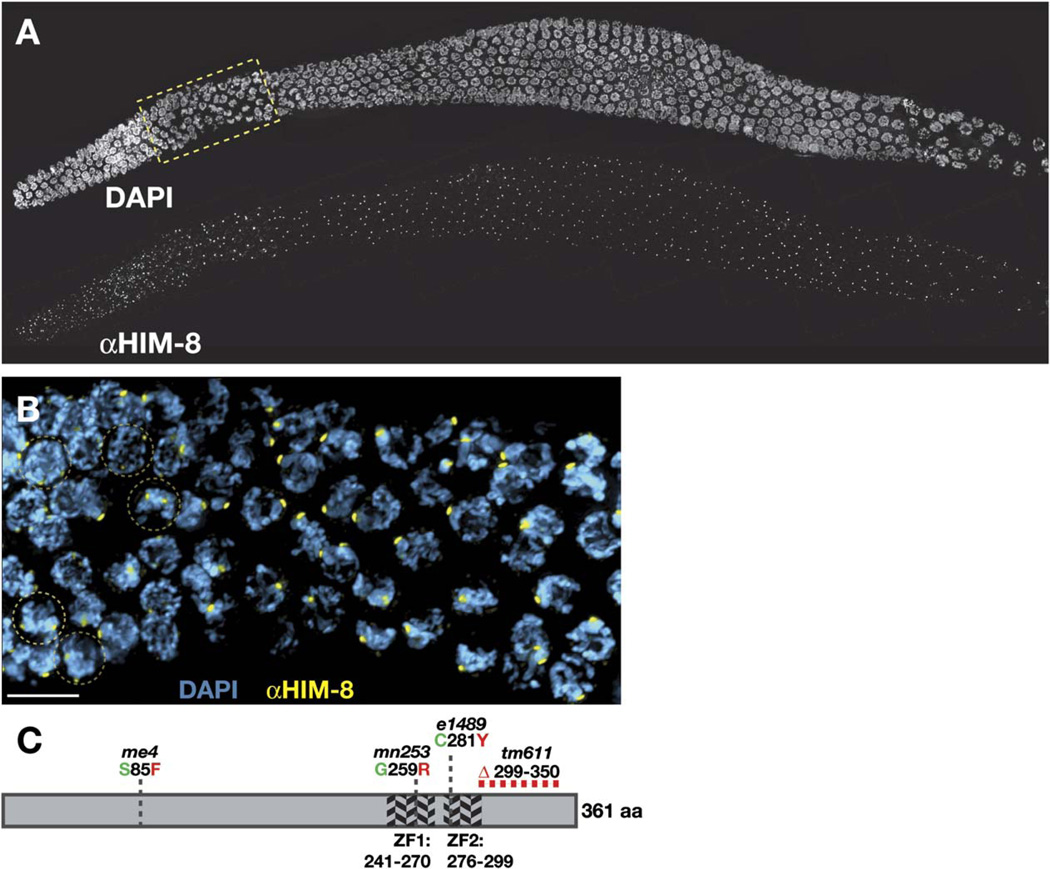
Figure 3. HIM-8 is a C2H2 Zinc-Finger Protein that Localizes to Distinct Nuclear Foci during Meiosis.
(A) Three-dimensional projection through a wild-type gonad stained with DAPI and antibodies against HIM-8. Subnuclear HIM-8 foci are present in all germ-line nuclei throughout the premeiotic, transition-zone, and pachytene region of the gonad. The transition-zone region outlined by the yellow box is magnified in (B).
(B) Prior to meiotic entry, two HIM-8 foci (yellow) are observed in each nucleus. Examples of premeiotic nuclei in which two foci can be clearly observed are outlined in brown circles. Once nuclei have entered the transition zone, representing the leptotene/zygotene stages of meiosis, they usually reveal only a single HIM-8 focus or closely spaced pair of foci. The scale bar represents 5 µm.
(C) Schematic representation of the HIM-8 protein. The diagram displays the location of two predicted zinc fingers as well as the sites of point mutations or deletions resulting from the four mutant alleles of him-8.

