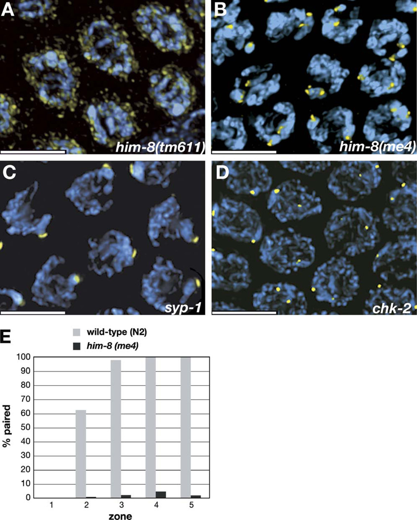
Figure 5. HIM-8 Localization in him-8 Mutants and Other Informative Meiotic Mutants.
All images show projections through the nuclear volumes of fields of pachytene-region nuclei from animals of the indicated genotypes. Immunofluorescence with anti-HIM-8 antibodies is shown in yellow, and DAPI staining is shown in blue.
(A) In him-8(tm611) mutant hermaphrodites, discrete foci of HIM-8 are not detected on the chromosomes. The intensity of the staining is shown more brightly here than in images of other genotypes to reveal that the residual staining is concentrated at the nuclear periphery. Similar staining is seen in him-8(e1489) and him-8(mn253) animals, which also carry mutations in the zinc-finger domain of HIM-8. This residual staining is detected using several different antisera raised against HIM-8, suggesting that it is specific rather than nonspecific background.
(B) In him-8(me4) hermaphrodites, two distinct foci are visible in each nucleus at pachytene. See also Figure 6B and Figure S1.
(C) In syp-1 hermaphrodites, a single focus of HIM-8 is detected in each nucleus in the pachytene region of the gonad, indicating that pairing of the HIM-8 binding region is stabilized despite the absence of synapsis.
(D) HIM-8 foci are detected on the X chromosomes but usually do not pair in chk-2 mutants.
(E) Immunofluorescence with the HIM-8 antibody was performed on wild-type and him-8(me4) hermaphrodites. The fraction of paired foci was scored in each of five zones of the gonad, which were defined in the same way as in our FISH analysis (Figure 2).
All scale bars represent 5 µm.

