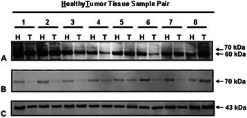
Fig. 5.

Immunoblotting of AChE in human upper airway epithelium. Proteins in non cancerous (Healthy; H) and cancerous (Tumour, T) pieces were separated by reducing SDS-PAGE and detected by western blotting with anti-AChE antibodies. The use of N19 antibodies allowed us to observe two-three deeply labelled protein bands of about 60-kDa and various fainter bands of 70–76 kDa (a). The active site-directed probe Ph-F was able to label the 70–76 kDa bands, but not the 60-kDa proteins (b). Accordingly, the deep 60-kDa protein bands in ANCT and HNSCC specimens were assigned to non-catalytic AChE proteins, and the faint 70–76 kDa bands to catalytic proteins. Note the much weaker signal corresponding to catalytic AChE in tumours (T) than healthy (H) tissues. The loaded control was β-actin (c)
