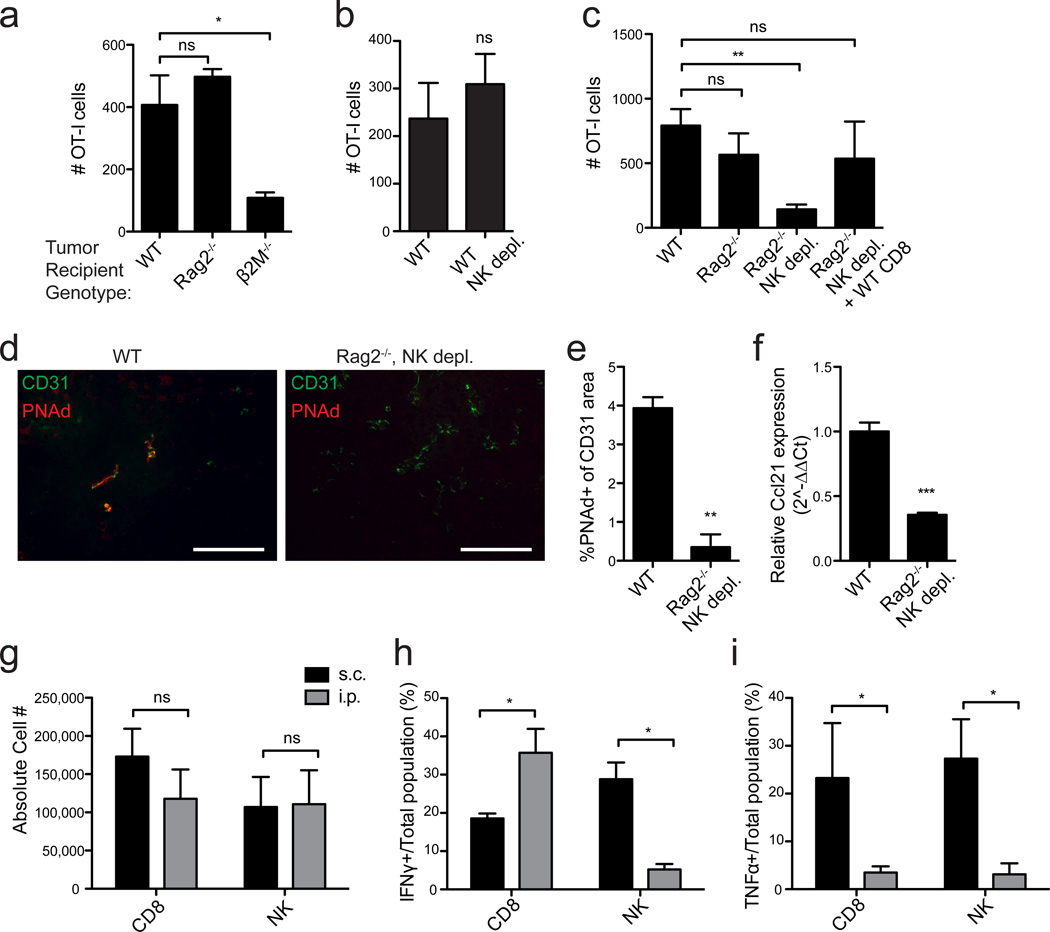
Figure 5. CD8 T-cells and NK cells act redundantly to induce LN-like vasculature in s.c. tumors.
(a–c) Infiltration of naïve OT-I cells into s.c. B16-OVA tumors in the indicated mice was determined as in Figure 2. n=3 (a) or 5–8 (b,c) per group.
(d,e) Representative images (d) and summary data (e, n=3) for PNAd expression in tumors in the indicated mice.
(f) Ccl21 expression (n=3) in s.c. tumor lysates was quantified as in Figure 3.
(g) Number of endogenous CD8 T-cells (CD3+CD8+) and NK cells (CD3−CD4−NK1.1+) present in s.c. (black bars) and i.p. (gray bars) tumors in WT mice was determined by flow cytometry. n≥8 per group.
(h,i) The percentage of total CD8 T-cells or NK cells producing IFNγ (h) or TNFα (i) in s.c. (black bars) or i.p. (gray bars) tumors from WT mice was assessed by intracellular staining 4 hr after Brefeldin A injection. n=3–5 per group
Data (mean+SEM) are representative of at least two independent experiments. ns:p>0.05, *p<0.05, **p<0.01, ***p<0.001 by unpaired t-test (b,e-i) or one way ANOVA with Dunnett’s post-test (a,c).

