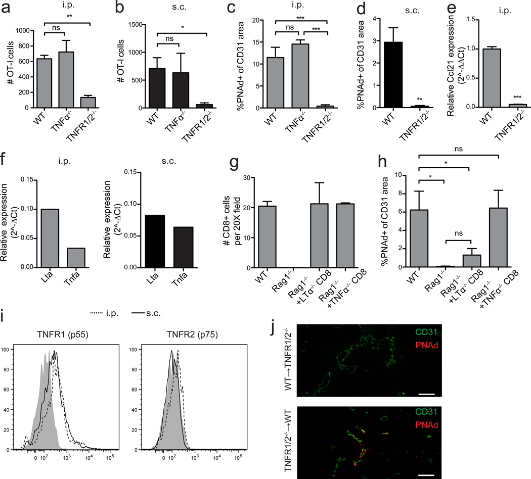
Figure 8. LTα3 induces PNAd expression on tumor vasculature by signaling through TNF receptors.
(a,b) Infiltration of naïve OT-I cells into i.p. (a) or s.c. (b) B16-OVA tumors in the indicated mice was determined. n=7 (a) or 4 (b) per group.
(c–e) PNAd expression (c,d) and Ccl21 expression in i.p. (c,e) or s.c. (d) tumors was quantified. n=4 per group.
(f) Expression of Lta and Tnfa mRNA in sorted CD3+CD8+ T-cells from i.p. (left panel) and s.c. (right panel) tumors was determined by quantitative RT-PCR. Expression levels are displayed relative to Hprt.
(g) Endogenous CD8 T-cell accumulation in d 14 i.p. B16-OVA tumors growing in WT, Rag1−/−, and Rag1−/− mice repleted with either TNFα−/− or LTα−/− CD8 T-cells mice (n=3 per group) was determined by immunofluorescence.
(h) PNAd expression in i.p. tumors from the indicated mice was determined. n=4 per group.
(i) Expression of TNFR1 and TNFR2 on CD31+ endothelial cells from i.p. (dashed lines) and s.c. (solid lines) B16-OVA tumors was determined by flow cytometry. Shaded region represents background staining in FMO controls.
(j) CD31 and PNAd expression in i.p. tumors from WT➔TNFR1/2−/− or TNFR1/2−/−➔WT bone marrow chimeras.
Data (mean+SEM) are pooled from two experiments (a,b) or representative of two (c–j) independent experiments. ns:p>0.05, *p<0.05, **p<0.01, ***p<0.001 by unpaired t-test (d–e) or one way ANOVA with Tukey’s post-test (a-c,h). Scale bars = 100 µm.

