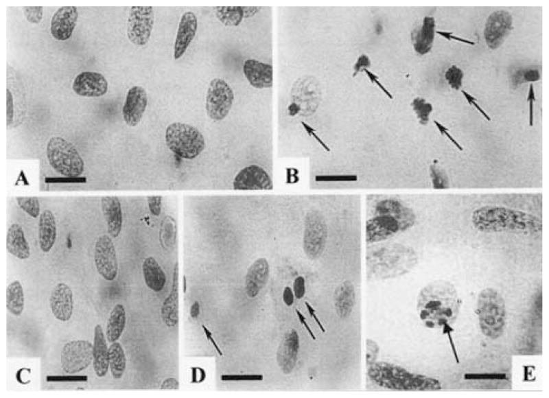
Fig. 2.
Morphology of nuclei of Feulgen-stained cultured cardiomyocytes after exposure to Ang II (10 nmol/L). (A) Nuclei of control cells. A few small granules against a pale background characterizes chromatin. (B) Exposure of myocytes to Ang II for 24 h led to an increase in the number of apoptotic nuclei. Condensation, compacting and margination of nuclear chromatin were accompanied by disappearance of the structural framework of the nucleus and nuclear breakdown. (C) Pretreatment with CCPA (1 μmol/ L) before Ang II. (D) Ang II treated cells, exposed to CPX (1 μmol/L) before pretreatment with CCPA (1 μmol/L). (E) Pretreatment with IB-MECA (0.1 μmol/L) before Ang II. Arrows indicate apoptotic cardiomyocytes. Bars = 10 μm.

