
Figure 3.
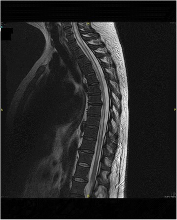
An abnormal moderately hyperintense on T2, hypointense T1, central intramedullary signal interesting almost all the cervical and dorsal spine respecting the terminal cone, and showing a contrast-enhanced periphery including a possible small necrotic remodeling over D7 and D8 with surrounding edema. The appearance is consistent with extensive myelitis, possibly infectious.