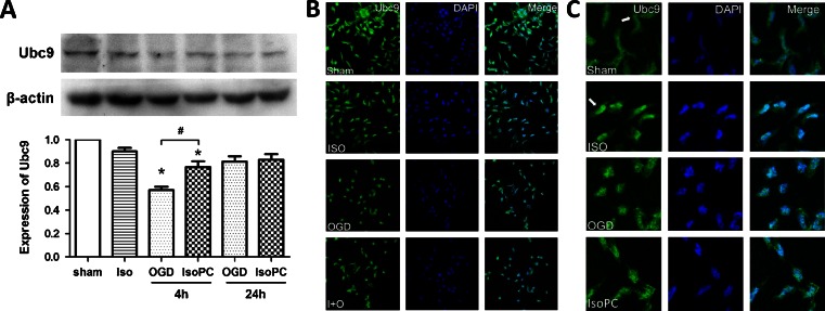
Fig. 2.
a Western blot results showing isoflurane preconditioning-induced recovery of Ubc9 expression hampered by OGD (n = 6 for each group). Data are presented as mean ± SEM. *p < 0.05 compared with the sham group. #p < 0.05 compared with the OGD 4-h group. b, c Representative immunofluorescent images for Ubc9 expression in different groups. Ubc9-positive cells were shown in green and cell nuclei were stained with DAPI (blue) (color figure online)

