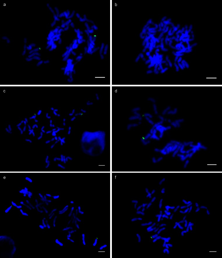
Fig. 1.
Fluorescence in situ hybridisation using probe UTV39 (green) on DAPI stained chromosomes of mitotic cells from root tip squashes of parental lines Brigand 8/2 (a) and Huntsman (b), translocation lines 12A5 (c), 12H3 (d) and deletion line 7C12 type 1 (e) and type 2 (f). Scale bar represents 10 µm

