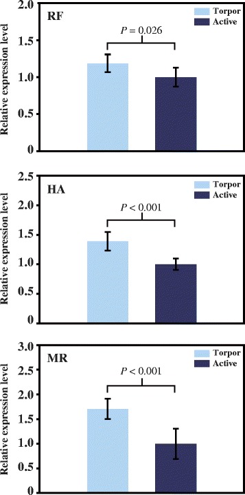
Fig. 2.

Expression levels of Pparα mRNA determined by real-time RT PCR. RF, HA, and MR represent hibernating R. ferrumequinum, H. armiger, and M. ricketti bats, respectively. Relative mRNA levels in torpid and active states are indicated with light blue and dark blue colors, respectively. The expression level of Pparα in active bats was set as 1.0. Data are presented as mean ± SD. A P value < 0.05 is considered statistically significant
