
Figure 1.
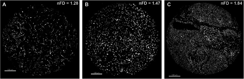
Representative DAPI-stained images of individual tissue microarray (TMA) cores used as substrates for nuclear fractal dimension (nFD) analysis. Image of an entire TMA core with (A) low nFD, (B) intermediate nFD and (C) high nFD.
Official websites use .gov
A
.gov website belongs to an official
government organization in the United States.
Secure .gov websites use HTTPS
A lock (
) or https:// means you've safely
connected to the .gov website. Share sensitive
information only on official, secure websites.
Representative DAPI-stained images of individual tissue microarray (TMA) cores used as substrates for nuclear fractal dimension (nFD) analysis. Image of an entire TMA core with (A) low nFD, (B) intermediate nFD and (C) high nFD.