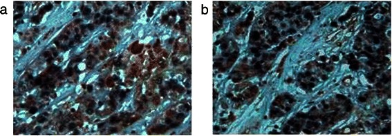
Figure 5.

Dual immunohistochemical analysis of apoptosis and TLR3 expression in HCC tissue. (a) In well-differentiated HCC tissue, the nuclei were TUNEL-positive and TLR3 was overexpressed at equal levels in the cytoplasm and membrane. (b) The nuclei of poorly differentiated HCC cells were TUNEL-positive, and these cells expressed relatively lower levels of cytoplasmic TLR3. Magnification × 400.
