
Figure 1. Multiple morphologies of MAM markers.
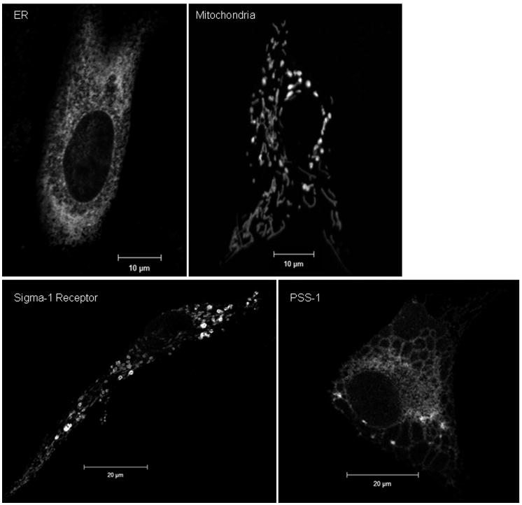
Representative morphologies of ER and mitochondrial organelles are shown in the top row. The characteristic reticular ER morphology was visualized using a commercially available, soluble ER lumen fusion protein, pECFP-ER (Clontech). Similarly, both the thread-like and punctuate morphologies associated with dynamic mitochondria are observed using a marker for the mitochondrial inner membrane, DsRed1-Mito (Clontech). Two different markers for the MAM are shown in the bottom row, Sig-1R (left panel) and PSS-1 (right panel), revealing multiple morphologies associated with this ER subdomain. Primary diploid fibroblasts were lipofected with either pECFP-ER, DsRed1-Mito, mEGFP-human PSS-1 [42], or with Sig1R-EYFP (a generous gift from Drs. Hayashi and Su) [14]. 24 hours after transfection, cells were fixed with ice cold methanol as described [42] and imaged using confocal microscopy with a Zeiss LSM510 and a 63× objective (NA 1.4). Panels show single optical sections (0.8 microns) of transfected cells.