
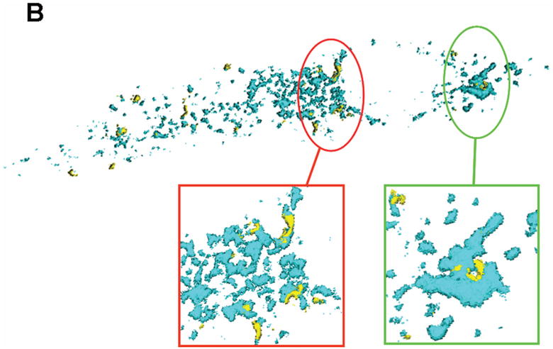
Figure 5. Physical proximity of HCMV pUL37x1 to lipid enriched MAM domains.
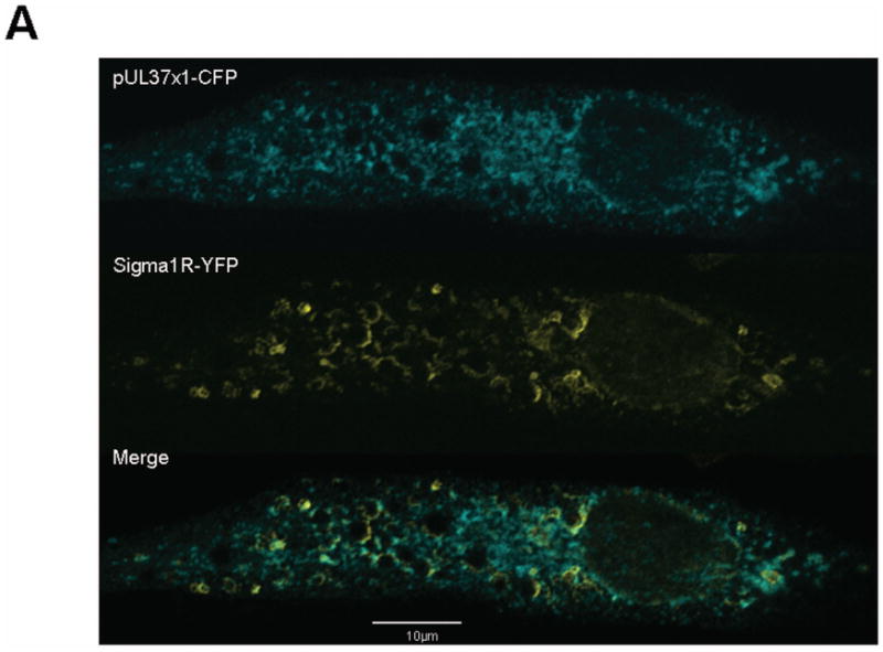
A. Shown is a single optical section of human fibroblasts expressing HCMV pUL37x1-Cerulean fluorescent protein (CFP) and Sig-1R-enhanced yellow fluorescent protein (EYFP). 24 hours after transfection, cells were fixed and imaged as in Figure 1.
B. Surface-rendered 3D recreation of a Z-stack series through the transfected cell in Panel A. Regions where pUL37x1 and Sig-1R are in close proximity are circled and enlarged on the bottom of the figure. Similar morphology and curvatures are seen with both proteins in these regions, suggesting that they occupy the same compartment.