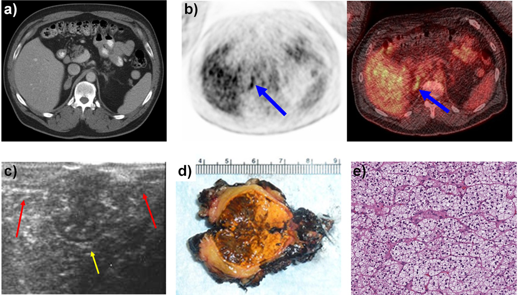
Figure 1.
a: CT Scan of 3 × 1.6 cm right Adrenal lesion with non-contrast enhancement of 14 HU
b: PET/CT Scan demonstrating an active foci with maximal SUV of 5.9 corresponding to the adrenal lesion.
c: Intra-operative ultrasound performed during right sided robotic partial adrenalectomy demonstrating normal adrenal limbs (red arrows) and a large central adrenal mass (yellow arrow).
d: Gross pathological examination of the bi-valved adrenal mass after right sided robotic partial adrenalectomy
e: Microscopic examination demonstrating adrenal pathology to be both micro and macronodular adrenal hyperplasia

