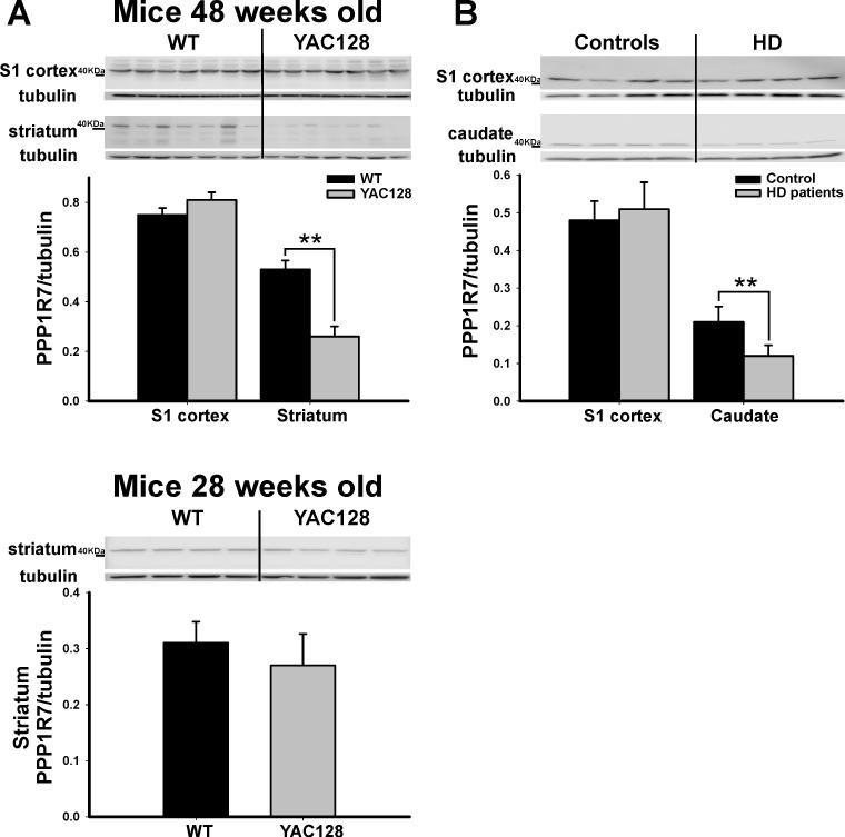
Figure 5. PPP1R7 deficiency correlates with anatomical phenotype.
(A) As shown in individual blots and by group data, PPP1R7 deficiency is observed in the striatum, but not the S1, in 48 weeks old YAC128 mice compared to WT littermates (48 weeks- upper panel). In contrast, 28 weeks old YAC128 mice compared to WT littermates showed no difference in PPP1R7 expression within the striatum (28 weeks- lower panel).
(B) As shown in individual blots and by group data, PPP1R7 deficiency is observed in the posterior body of the caudate, but not the S1, in HD human brains compared to controls. Lower panels in A and B show PPP1R7/Tubulin mean ratios and SE.
* denotes p≤0.05, ** denotes p≤0.01

