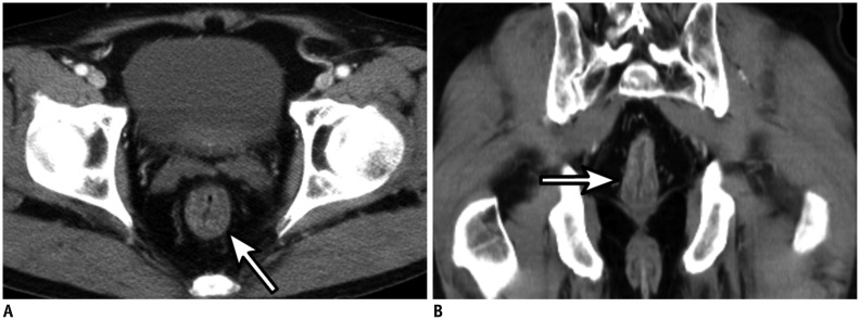
Fig. 3. 85-year-old male patient with massive hematochezia.
A, B. Axial arterial phase (A) and coronal portal venous phase (B) CT angiography showing potential bleeding focus at rectum. Note edematous wall thickening of segmental rectal wall (arrow). Subsequent colonoscopy revealed hyperemia on mucosal layer of rectum which was considered potential bleeding focus.

