
Fig. 7. Ventral perspective of three-dimensional reconstruction of MR images of photothrombotic cerebral infarction and superparamagnetic iron oxide (SPIO)-labeled human bone marrow-derived mesenchymal stem cells (hBM-MSCs) in rats.
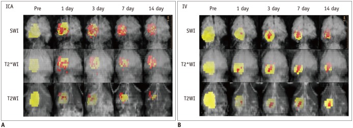
Color yellow was assigned to cerebral infarction, and color red was assigned to SPIO-labeled hBM-MSCs surrounding infarction. Internal carotid arterial (ICA) injection (A) of SPIO-labeled hBM-MSCs shows engraftment of more cells in earlier days than intravenous (IV) injection (B). SWI = susceptibility-weighted images, T2WI = T2-weighted images, T2*WI = T2*-weighted images