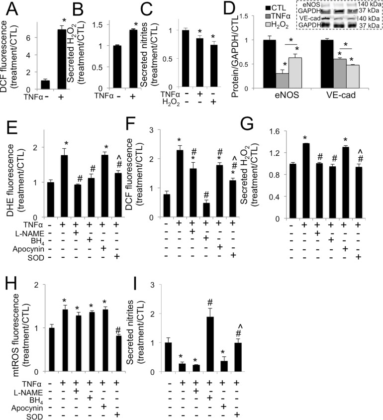
Fig 3 is incorrect in panels E through I. The authors have provided a corrected version here.
Fig 3. TNFα drives increased oxidative stress in aortic valve endothelial cells via eNOS uncoupling.
A, TNFα increases oxidative stress in VEC at 30 minutes. B, TNFα increases hydrogen peroxide (H2O2) secretion from VEC at 30 minutes. C, TNFα or H2O2 decrease nitric oxide secretion from VEC at 48 hours (n = 4). D, TNFα or H2O2 decrease eNOS and VE-cadherin expression in VEC at 48 hours. Representative western blot images (inset) and blot quantification. E, L-NAME, BH4, or peg-SOD but not apocynin block increases in superoxide (DHE) in VEC caused by TNFα, at 30 minutes. F, L-NAME, apocynin, and peg-SOD mitigate increases in general oxidative stress (DCF) caused by TNFα at 30 minutes, but only BH4 completely blocks superoxide increase, maintaining control levels. G, L-NAME, BH4, or peg-SOD but not apocynin block increases in H2O2 secreted by VEC at 30 minutes caused by TNFα at 30 minutes. H, TNFα drives increased mtROS, mitigated only by co-treatment with SOD. I, BH4, or peg-SOD but not L-NAME or apocynin block decreases in nitric oxide secretion in VEC caused by TNFα at 48 hours. * indicates p < 0.05 versus control. # indicates p < 0.05 versus TNFα. ^ indicates p < 0.05 versus apocynin. N = 4.
Reference
- 1. Farrar EJ, Huntley GD, Butcher J (2015) Endothelial-Derived Oxidative Stress Drives Myofibroblastic Activation and Calcification of the Aortic Valve. PLoS ONE 10(4): e0123257 doi: 10.1371/journal.pone.0123257 [DOI] [PMC free article] [PubMed] [Google Scholar]


