
FIG 4 .
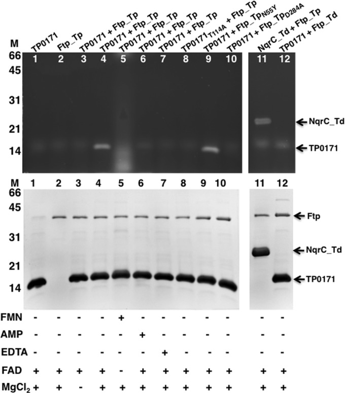
SDS-PAGE characterization of flavinylation reactions followed by UV illumination and Coomassie blue staining. UV illumination of unstained gel is shown at the top, and the Coomassie-stained gel is shown below. Protein molecular markers are on the left side. Ftp_Tp (wild type and mutants) reacted with TP0171 (wild type and mutants) and NqrC_Td under various indicated conditions. The nonspecific diffuse bands (observed in all lanes, including the control reactions) are in vitro artifacts of the flavinylation reactions.