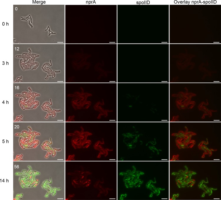
FIG 3.
nprA and spoIID promoter activities in cells developing in a microcolony. Cells of the spoIID-yfp nprA-mCherry-expressing Bt strain were allowed to grow at 30°C on a solid medium containing LBP diluted 1:100, and mCherry and YFP expression was monitored by time-lapse microscopy. The fluorescent mCherry and YFP signals were false colored red and green, respectively. Snapshots are taken from Movie S1 in the supplemental material. Time zero is the first frame of the movie. The numbers in the left panels are frame numbers. Merge, merged image of the signals in the fluorescent channels and the phase-contrast image; nprA, mCherry fluorescence associated with the activity of the nprA promoter; spoIID, YFP fluorescence associated with the activity of the spoIID promoter; overlay, overlay of the signals in both fluorescent channels. Bars, 10 µm.

