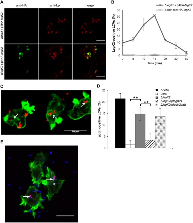
FIG 4 .
LegK2 inhibits actin polymerization on the LCV. (A) HA-LegK2 detection on LCVs. D. discoideum was infected for 15 min at an MOI of 100 with dotA or legK2 mutant L. pneumophila transformed with a vector encoding N-terminally HA-tagged LegK2. The presence of bacteria was detected by an immunofluorescence assay with anti-MOMP antibodies (anti-Lp, red labeling), and the HA-LegK2 fusion is labeled with anti-HA antibodies (anti-HA, green labeling). Scale bars, 5 µm. (B) HA-LegK2 detection during LCV biogenesis. D. discoideum was infected at an MOI of 100 with legK2 mutant L. pneumophila transformed with a vector encoding N-terminally HA-tagged LegK2. HA-LegK2-positive LCVs were counted at 0, 5, 10, 15, 20, 30, and 60 min after bacterium-amoeba contact (>100 LCVs per time point). These data are representative of three independent experiments, and the error bars represent the standard deviations. (C) Actin polymerization on LCVs during Legionella infection. D. discoideum was infected for 15 min at an MOI of 100 with mCherry-labeled legK2 mutant L. pneumophila. Polymerized actin on LCVs was detected by labeling with phalloidin-FITC. Arrows show examples of actin-positive LCVs. Scale bar, 10 µm. (D) Detection of polymerized actin in legK2 mutant-containing vacuoles. Actin-positive vacuoles (n = >100) were counted for amoeba infected with WT L. pneumophila strain Lens, the derivative dotA and legK2 mutants, and the transformed legK2(plegK2) and legK2(plegK2cat) mutants. These data are representative of three independent experiments, and the error bars represent the standard deviations. **, P < 0.01. (E) Exclusive detection of LegK2 and actin on LCVs. D. discoideum was infected for 15 min at an MOI of 100 with L. pneumophila transformed with a vector encoding HA-tagged LegK2. Bacteria were detected with anti-Lp1 serum and an anti-rabbit secondary antibody (blue labeling), HA-LegK2 on the LCVs was labeled with an anti-HA antibody and an anti-mouse secondary antibody (red labeling), and actin was stained with phalloidin-FITC (green labeling). Arrows indicate LegK2-positive LCVs (without actin labeling), and the dotted arrow indicates an LCV without LegK2 on its surface that was consequently stained for actin. Scale bar, 10 µm. This micrograph is representative of 50 LCVs observed in two independent experiments.

