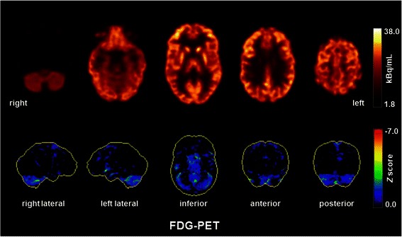
Figure 2.

[18F]fluorodeoxyglucose positron emission tomography (FDG-PET) showed a mild, borderline significant cerebellar hypometabolism. Otherwise, the PET was unremarkable. No mesiotemporal hypermetabolism as typically seen in limbic encephalitis was observed. FDG-PET was performed at the Department of Nuclear Medicine of the University Hospital Freiburg after injection of 320 MBq FDG (Gemini True Flight, Philips Electronics, The Netherlands). Upper and lower row images show transaxial FDG-PET images and 3D surface projections of regions with decreased FDG uptake (colour-coded Z-score, compared to age-matched healthy controls), respectively.
