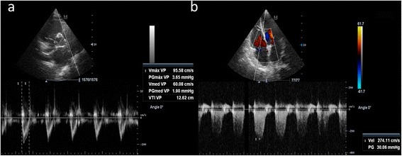
Fig. 1.

Example for estimating the pulmonary vascular resistance. a. Right ventricle outflow tract velocity time integral (12.6 cm) obtained in parasternal short-axis view. b. Right ventricle to right atrial peak velocity (2.7 m/s) obtained from a tricuspid regurgitation jet in apical four-chamber view. PVR is obtained as (2.7 /12.6) × 10 + 0.16, equal to 2.3 Wood units (normal <1.5)
