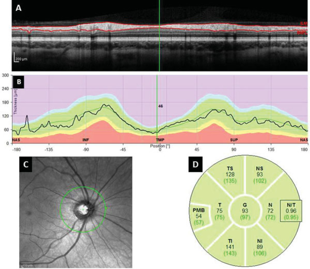
Fig. 2.
SD–OCT image of the Circumpapillary Retinal Nerve Fiber Layer Thickness Measures. (A) Single B-scan of the circumpapillary SD–OCT acquisition. Solid red lines demonstrate automated segmentation of the RNFL. (B) RNFL thickness measures of the OCT image at anatomic locations. (C) Near-infrared image demonstrating en face view of the 3.45 mm circle centered over the optic nerve. (D) Quadrant, subquadrant, and global average RNFL thickness measures. N, nasal; NI, nasal inferior; NS, nasal-superior; PMB, papillomacular bundle; SD–OCT, spectral domain optical coherence tomography; T, temporal; TS, temporal-superior; TI, temporal inferior.

