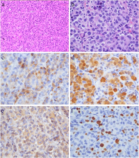
Figure 2.

Pathologic results. (a) Pathology slice (hematoxylin-eosin stain, original magnification ×100) of the tumor. Nests of intermediate-grade tumor cells with intervening stroma can be seen. The tumor cells show clear to eosinophilic cytoplasm, with no obvious pigment. (b) Higher magnification (hematoxylin-eosin stain, original magnification ×400) of the tumor, showing prominent nuclear mitosis and atypia. (c) Higher magnification (original magnification ×400) of the tumor, showing HMB45 positive. (d) Higher magnification (original magnification ×400) of the tumor, showing S-100 positive. (e) Higher magnification (original magnification ×400) of the tumor, showing Vimentin-positive. (f) Higher magnification (original magnification ×400) of the tumor, showing Ki-67 index, was 20% to 30%.
