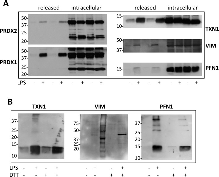
Fig 1. LPS induces release of PRDX1, PRDX2, TXN1, VIM and PFN1.
Western blot analysis following non-reducing (A) or reducing (B) SDS-PAGE (10% acrylamide for VIM, 12% for PRDXs, 15% for TXN1 and PFN1) of RAW264.7 supernatants cultured with and without 100 ng/ml LPS for 24 h. Supernatants were blocked with 40 mM NEM immediately after collection, to prevent thiol-disulfide exchange. Cell lysates were also analyzed after blocking with NEM.

