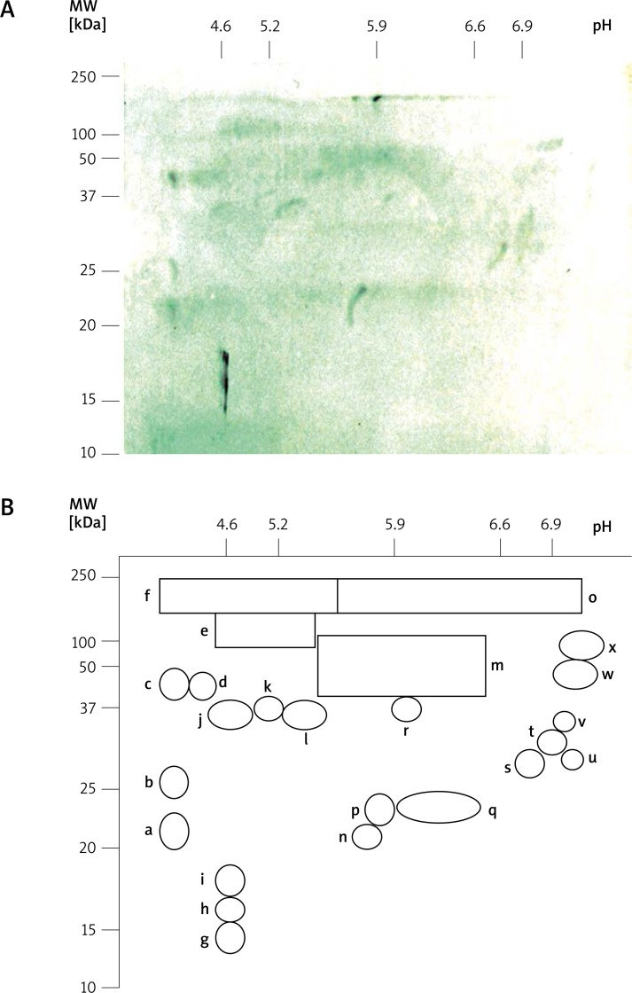
Figure 3.
A – IgE immunoblot with a pool of sera from 2-D Der p separation (dog Der p allergogram), revealed with horseradish peroxidase-conjugated goat anti-dog-IgE. B – Two-dimensional diagram from dog Der p allergogram in Figure 3A, with the recognized spots identified from a to x

