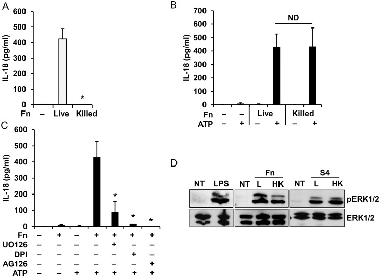
Fig 5. Monocyte priming by Francisella is ERK dependent.
Human monocytes were infected for 16 h with live or killed F. novicida (Fn) and IL-18 release was measured by ELISA (A). Human monocytes were primed with live or killed F. novicida for 30 min followed by ATP stimulation for 30 min and release of IL-18 was measured by ELISA (B). IL-18 release from monocytes pretreated with UO126 (20 μM), AG126 (10 μM) or DPI (50 μM) for 30 min and then primed with F. novicida (Fn) for 30 min followed by ATP stimulation for 30 min (C). Monocytes, primed with live (L) or heat killed (HK) F. novicida (Fn) and F. tularensis SchuS4 (S4) for 30 min were lysed and cell lysate was analyzed for ERK phosphorylation. LPS from E. coli was used as a positive control (D). Data represent mean ± SEM, n = 3 independent experiments. * p<0.05. ND—not statistically different. Blots are representative of repeated experiments. White bars—overnight model, black bars—rapid priming model.

