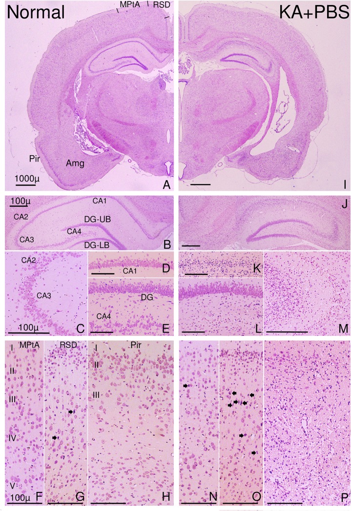
Fig 3. Photomicrographs of the forebrain of a normal rat (left half) or a KA and PBS-injected rat (right half) with severe damage in the hippocampus and piriform (Pir) cortex.
Many neurons in CA1-4 were damaged in the hippocampus of the rat injected with KA (J–M). In the cortex of the rat injected with KA (N–P), few neurons were damaged in the medial parietal association cortex (MPtA), some neurons were damaged in the retrosplenial dysgranular cortex (RSD; O), and many neurons were damaged in the piriform cortex (P). Damaged neurons are indicated by closed arrows. Also, some dead neurons were detected in the RSD of normal rats (G). DG-UB, DG-LB: Upper and lower blade of the dentate gyrus (DG). Scale bar = 1,000 μm (A, I); scale bar = 100 μm (B–H, J–P).

