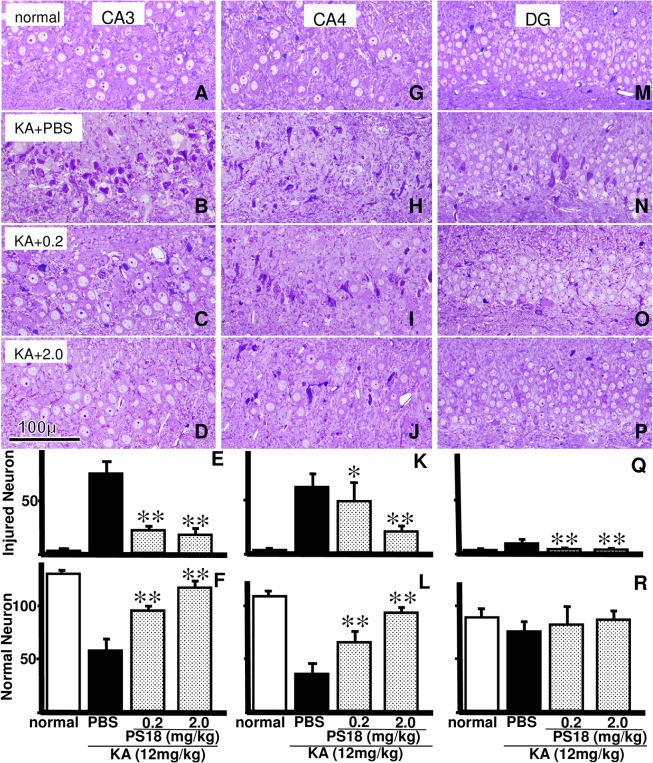
Fig 7. Light Microscopic Analysis of Other Hippocampal Regions.
Photomicrographs of toluidine blue-stained hippocampal neurons in CA3 (A-D), CA4 (G–J), and the upper blade of the dentate gyrus (DG; M–P) in normal control rats (A, G, M) and rats that received an injection of PBS (B, H, N), 0.2 mg/kg PS18 (C, I, O), or 2.0 mg/kg PS18 after a KA injection (D, J, P). Injured CA1 neurons were rescued by the PS18 treatment. Note that some neurons were also injured in the DG (N). Scale bar = 100 μm. Effects of PS18 on injured (E, K, Q) and normal (F, L, R) neuronal density of the hippocampal CA1 region in rats that received a subcutaneous injection of 12 mg/kg kainic acid (KA). PS18 treatment following a KA injection in rats decreased the number of injured neurons (E, K, Q) and increased the number of viable neurons (F, L, R) in a dose-dependent manner compared with PBS-treated KA-injected rats. *P < 0.05, **P < 0.01. A P-value < 0.05 was considered to be statistically significant. All data are expressed as mean ± standard error of the mean (S.E.M.).

