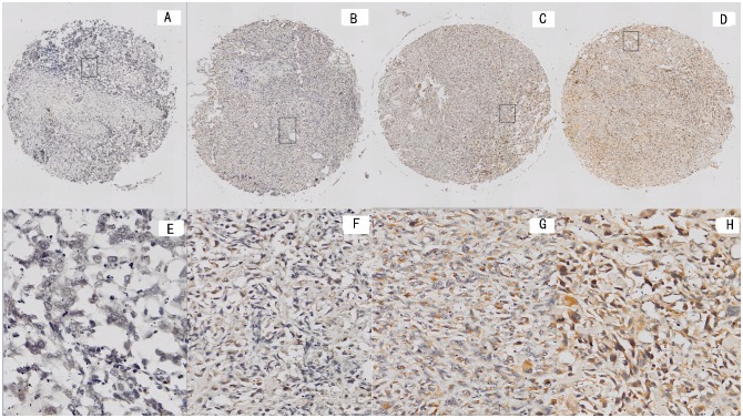
Fig 2. Representative immunohistochemical staining forPLA2G16 in tissue microarray.
Representative PLA2G16 staining samples at magnification of 40at levels of 0, 1, 2, and 3 (A), (B), (C) and (D).RepresentativePLA2G16 staining samples at magnification of 200 at levels of 0, 1, 2, and 3 (E), (F), (G) and (H).

