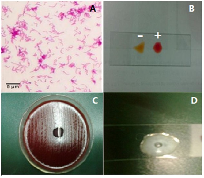
Fig 2. Identification of H. pylori.

A: Gram staining, Gram staining differentiates bacteria by detecting peptidoglycan, which exists in the cell walls of gram-positive bacteria. After a Gram stain test, gram-positive bacteria shows the crystal violet dye, while a counterstain (safranin) added, gives gram-negative bacteria a pink coloring. B, rapid urease test, is a rapid diagnostic test for H. pylori. H. pylori secrete the urease enzyme, which can catalyze the conversion of urea to NH3 and raises the pH of the medium. The medium contains urea and an indicator such as phenol red, and thus the raised pH changes the color of the medium from yellow (a blank control) to red (an experimental group). C, the oxidase test is used to determine if a bacterium produces certain cytochrome c oxidases. It uses disks impregnated with a reagent such as TMPD, which is a redox indicator. The reagent can be changed from a dark-blue to maroon color when it is oxidized. D, the catalase test is one of main tests to identify H. pylori. The presence of catalase enzyme in the species is detected via hydrogen peroxide. If the bacteria possess catalase, when bacterial isolate is added to hydrogen peroxide, bubbles of oxygen will be observed. If the mixture produces bubbles, the organism is regarded as 'catalase-positive'.
