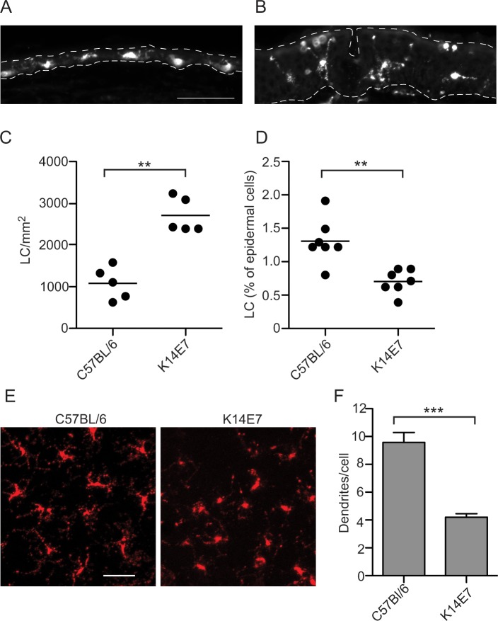
Fig 1. The absolute number of LC is increased in K14E7 mouse epidermis.
Sections of C57Bl/6 (A) and K14E7 (B) mouse ear were stained with CD207 and imaged by epifluorescence microscopy. The epidermis (top) is indicated by a broad dashed line, and the epidermis/dermis interface by a narrow dashed line; scale bar = 50μm. LC (CD45.2, CD11c, CD207, MHCII+) harvested from a 5.5 mm diameter sample of dorsal ear epidermis were enumerated, with counting beads, by flow cytometry. The absolute number (C) and the percentage of LC (D) are shown. Confocal imaging was carried out on CD207-stained sheets of dorsal ear skin (E). Scale bar represents 20μm. Dendrites on the CD207+ LC were enumerated following confocal imaging (F). **P < 0.01 (Mann-Whitney U test).

