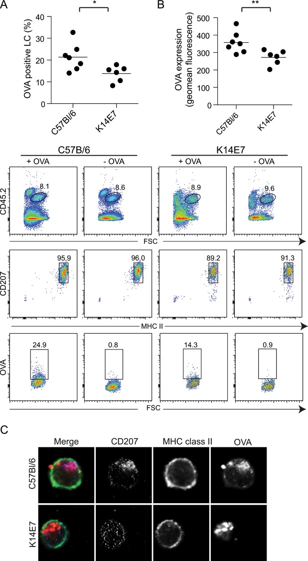
Fig 2. Antigen uptake is reduced in K14E7 mice.
Epidermal cell suspensions from C57BL/6 and K14E7 mice were cultured with Alexa-555-labelled OVA overnight at 37°C, stained and analysed by flow cytometry (A). The Alexa-555-OVA+ LC (CD45.2+, MHC Class II+, CD207+) as a percentage of total LC (B) and the level of expression of Alexa-555-OVA on the positive stained LC is shown (C). **P < 0.01; *P < 0.05 (Mann-Whitney U test). Confocal imaging of CD207+, MHCII+ LC that have taken up Alexa-555-OVA (D).

