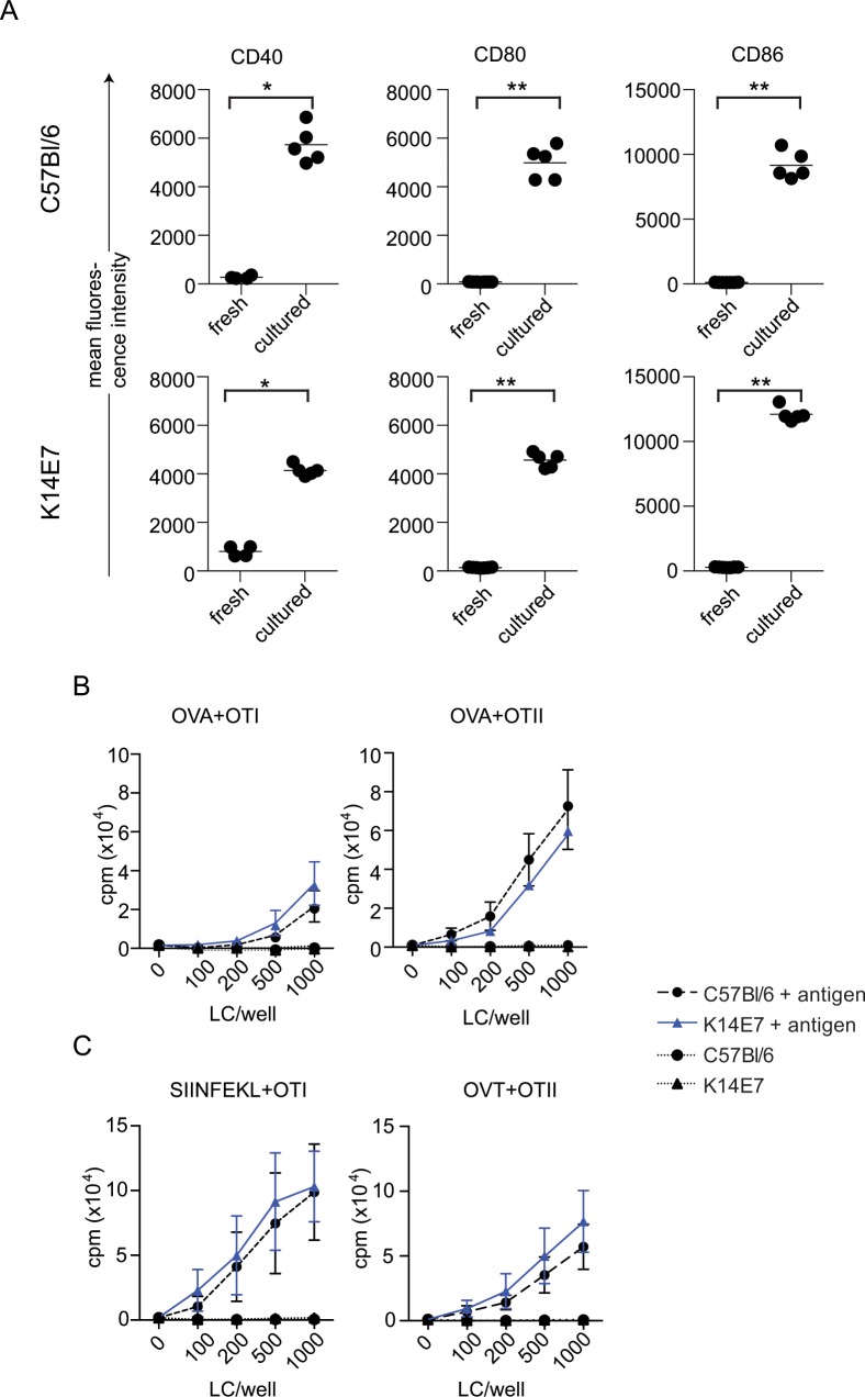
Fig 6. Co-stimulatory molecule expression is increased, and presentation and cross-presentation by LC from K14E7 mice does not differ from LC from C57Bl/6 mice.
Expression of CD40, CD80 and CD86 was compared on CD45.2, CD11c, CD207 positive LC from C57Bl/6 and K14E7 mice prepared either directly from epidermal suspensions (fresh) or from epidermal cells that were matured in culture for 72 h in medium containing GM-CSF (A). Epidermal cells from C57BL/6 and K14E7 mice were incubated with OVA, cultured for 2 days then LC purified and co-cultured with purified OT-I or OT-II cells for 60 h. T cell proliferation was measured after addition 3H-thymidine for a further 16h (B). Epidermal cells were cultured for 72 h in GM-CSF, LC purified and pulsed with SIINFEKL or ISQAVHAAHAEINEAGR for 2 h, washed and co-cultured with T cells for 60 h. T cell proliferation was measured after addition 3H-thymidine for a further 16h (C). Data was analysed using 2-way ANOVA and there was no significant difference between the groups.

