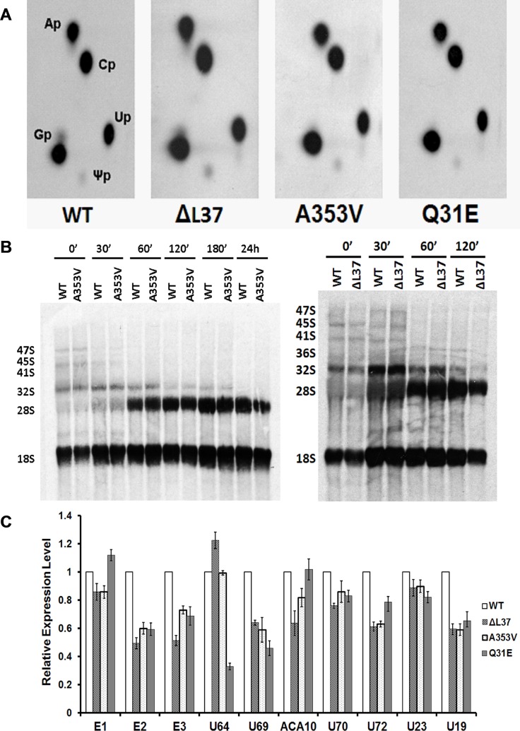
Fig 2. DKC1 mutant iPS cells show no significant defects in ribosome biogenesis.
A: measurement of pseudouridine in 28S rRNA from WT and mutant iPS cells. iPS cells were labeled with 32P-labeled orthophosphate and 28S rRNA was gel-purified. After digestion with RNase T2, each sample was separated by two-dimensional TLC. The positions of the labeled ribonucleotides are indicated. Ap: Adenine, Cp: Cytosine, Gp: Guanine, Up: Uridine, Ψp: Pseudouridine: B: Pulse–chase labeling experiments of rRNA isolated from WT, A353V and ΔL37 iPS cells. Cells were labeled with L-[3H-methyl] methionine for 30 min and then chased in nonradioactive medium for the times shown. The RNA was separated on a 1.25% agarose gel, transferred to a nylon filter, and exposed to x-ray film. C: Real-time RT/PCR results of some H/ACA snoRNAs of WT and DKC1 mutant iPS cells. Results were expressed relative to GAPDH RNA. The combined results of 3 independent experiments are shown, the error bars show standard deviation.

