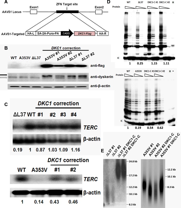
Fig 3. Expression of WT Dyskerin can rescue dysfunctional telomerase in ΔL37 cells but not in A353V cells.
A: Schematic representation of zinc-finger nuclease (ZNF)-mediated homologous recombination. The constitutively active AAVS1 “safe harbor” locus is shown on the top line and the targeting construct is shown below. The cDNA expression cassettes driving expression of Flag-tagged WT dyskerin under the chicken actin promoter (CAGG) were inserted by zinc finger-mediated homologous recombination into intron 1 of AAVS1. HA, homologous arms left (L) and right (R); SA-2APuro-PA, puromycin drug resistance cassette. [30] B: Western blot showing expression of flag-tagged dyskerin protein in corrected A353V and ΔL37 iPS cells by using anti-Flag antibody and anti-dyskerin antibody. Open arrow: endogenous dyskerin protein; filled arrow: Flag-tagged WT dyskerin protein. C: Northern blot result of TERC RNA expression levels of DKC1 corrected iPS cells. Different corrected lines are shown as well as the uncorrected line carrying the DKC1 ΔL37 or the DKC1 A353V mutation. β-actin was used as loading control. The quantitive data, derived by densitometry, are shown. D: The TRAP assay was performed to measure the telomerase activity after expressing the WT DKC1 gene in A353V and ΔL37 iPS cells, uncorrected and corrected by ectopic expression of WT DKC1 (DKC1-C). The quantitive data, derived by densitometry, are shown E: Telomere lengths of DKC1 corrected iPS cells were measured by using in-gel hybridization with a telomere probe (TTAGGG)3. DKC1-C indicates corrected iPS cells expressing WT DKC1 gene.

