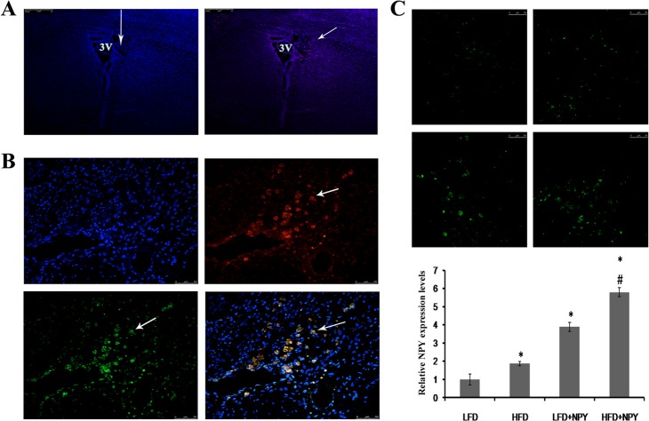
Fig 1. Constitutive overexpression of NPY in the paraventricular nucleus (PVN) of rats.
Eight weeks after the LV-Cherry (vehicle control) or LV-NPY-Cherry injection, the expression of the vectors was measured using immunofluorescence. 2 rats were excluded because of imprecise injection. (A) The lentiviral vectors LV-Cherry or LV-NPY-Cherry were injected vertically into the PVN (left panel). The arrow shows the direction of injection and the position of the PVN in the rat brain, and the blue immunofluorescence indicate nucleus. Expression of the reporter protein Cherry (red) in PVN after injection of LV-Cherry (right panel). The arrow indicates Cherry expression in neurons. 3V: the third ventricle of cerebrum (amplification: 100x). (B) Photomicrographs of NPY and Cherry overexpression in PVN after lentivirus injection. blue immunofluorescence for nucleus, green for NPY, red for cherry, and yellow for co-localization of NPY and cherry (amplification: 400x). The arrows indicate NPY and/or Cherry expression in a single neuron of the PVN. (C) The representative images of NPY overexpression from LFD (top left), HFD (top right), LFD+NPY (left bottom) and HFD+NPY (right bottom) groups, and quantification data is shown in the bottom (n = 8) (amplification: 400x). Data are presented as means ± SEM, *P<0.5 vs. LFD; # P<0.5 vs. HFD.

