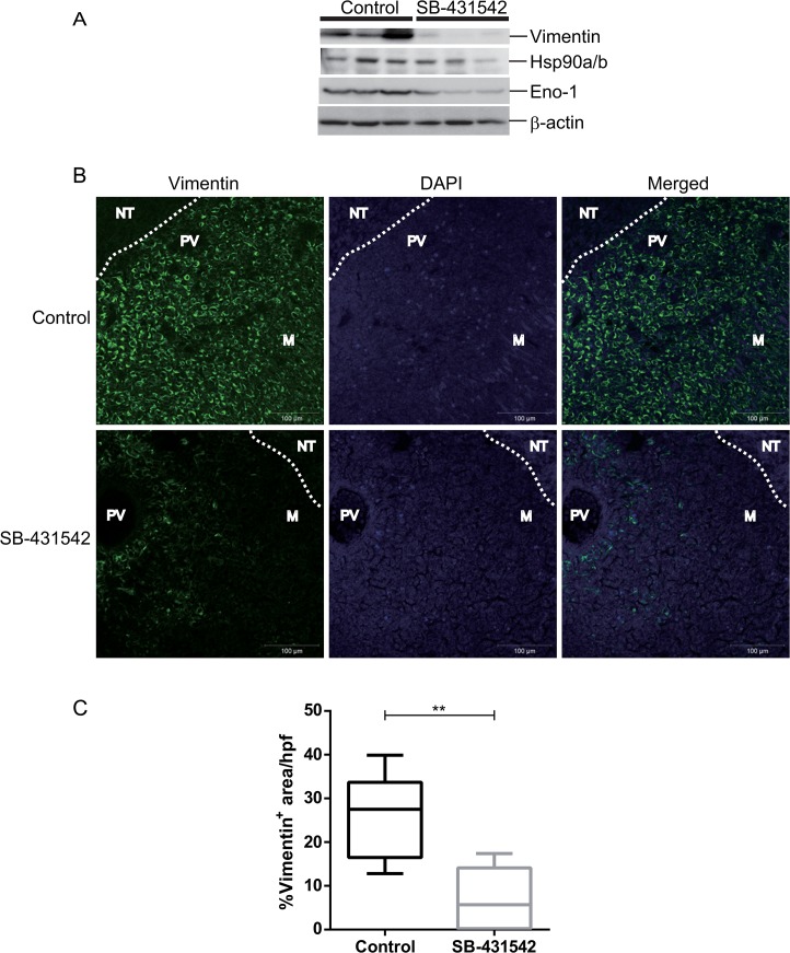
Fig 5. Validation of protein expression using western blot and immunohistochemistry.
(A) Metastases lysates were prepared from three mice per group and evaluated by western blot with anti-vimentin, anti-Hsp90a/b, and anti-Eno1 antibodies. β-actin was used as internal normalization. (B) Representative image of positive vimentin staining is shown exclusively in the metastases lesion and not in the surrounding lung tissue. White dotted lines represent a boundary of tumor and surrounding normal lung tissue. M, metastases; NT, normal lung tissue; PV, pulmonary vein. (C) Randomly selected high power fields were immunostained with vimentin and were quantitated using Zeiss software with the % area occupied by metastasized nodules in 4T1 metastasized tumors. A significant reduction in the number of vimentin positive cells was seen in the SB-431542-treated tumors. Scale bars represent 100 μm; hpf, high power fields.

