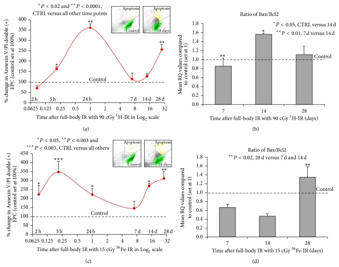
Figure 6.
Full-body 1H-IR and 56Fe-IR induces early (2–24 h) and delayed (14–28 days) apoptosis in BM-EPCs ex vivo. Graphic representation of mean % change in Annexin V and propidium iodide (P.I) double positive (+) BM-EPCs cultured ex vivo for 72 h (solid red line) after full-body single-dose IR of (a) 90 cGy 1H-IR mice and (c) 15 cGy 56Fe-IR mice at 2, 5, and 24 hours and 7, 14, and 28 days after IR. The corresponding control for each time point was set at 100%. Insets in (a) and (c) are representative flow cytometry analysis plots for corresponding control and 28-day time points. Graphic representation of qRT-PCR analysis, mean RQ values compared to control (which was set at 1) of BM-EPCs from full-body single-dose IR of (b) 90 cGy 1H-IR mice and (d) 15 cGy 56Fe-IR mics at 7, 14, and 28 days after IR for ratio of Bax/Bcl2. The corresponding control for each time point was set at 1. Graphs represent mean ± SEM of the pooled data from 5-6 independent biological samples/experiments. Statistical significance was assigned when P < 0.05.

