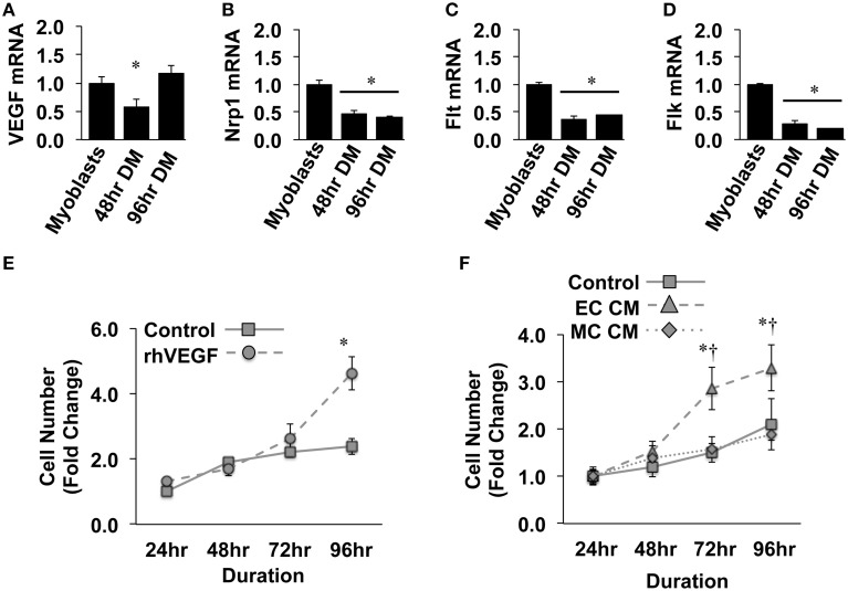
Figure 1.
Ischemic endothelial cells promote muscle precursor cell (MPC) proliferation. (A–D) Muscle myoblasts were grown to confluence and placed in differentiation medium (DM) for 48- or 96-h. QRT-PCR was used to determine the mRNA expression levels of VEGF (A), Neuropilin/Nrp1 (B), VEGFR1/Flt (C), and VEGFR2/Flk (D). (E) Primary mouse MPCs were plated at approximately 20% confluence, treated with recombinant VEGF (50 ng/mL), and cell number was determined at the indicated time points. (F) Primary mouse MPCs were plated at approximately 20% confluence, treated with conditioned medium from muscle cells (MC CM) or HUVECs (EC CM) that had been subjected to experimental ischemia (HND), and cell number was determined at the indicated time points. Cell proliferation rates were normalized to Control values at 24-h post-plating. *P < 0.05 vs. Myoblasts (QRT-PCR) or time-matched control (proliferation). †P < 0.05 vs. time-matched MC CM treatment group. All values are means ± SE.

