Figure 3.
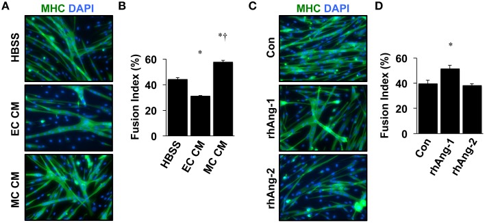
Ischemic myotubes and Ang-1 promote myoblast differentiation. (A) Representative immunofluorescently labeled images of differentiated myotubes (96 h) for visualization of myosin heavy chain (MHC, green) and nuclei (DAPI, blue) after treatment in differentiation medium supplemented with 25% by volume vehicle (HBSS) or conditioned medium from experimentally ischemic HUVECs (EC CM) or myotubes (MC CM). (B) Quantification of the percentage of nuclei incorporated into multinucleated myotubes (Fusion Index, %) after CM treatment. (C) Representative images of immunofluorescently labeled differentiated myotubes (96 h) after treatment in differentiation medium supplemented with Ang-1 (500 ng/mL), or Ang-2 (500 ng/mL) Green: myosin heavy chain (MHC); blue: DAPI. (D) Quantification of the percentage of nuclei incorporated into multinucleated myotubes (Fusion Index, %) after angiopoietin treatment. *P < 0.05 vs. Control or Myoblast, †P < 0.05 vs. EC CM treatment group. All values are means ± SE.

