Figure 4.
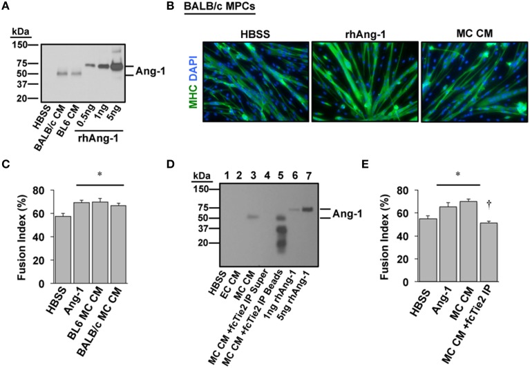
Angiopoietin-1 is secreted by ischemic myotubes to promote muscle cell differentiation. (A) Western blotting of CM from primary myotubes derived from ischemia-susceptible (BALB/c) and ischemia-resistant (BL6) parental strains verifies the presence of Ang-1 protein. (B) Representative images of primary BALB/c muscle progenitor cells differentiated into myotubes (96 h) after treatment in differentiation medium supplemented with vehicle (HBSS), Ang-1 (500 ng/mL), or conditioned medium from experimentally ischemic BALB/c myotubes (MC CM). Green: myosin heavy chain (MHC); blue: DAPI. (C) Quantification of the percentage of nuclei incorporated into multinucleated myotubes (Fusion Index, %) after treatment in differentiation medium supplemented with vehicle (HBSS), Ang-1 (500 ng/mL), or conditioned medium from experimentally ischemic BL6 or BALB/c myotubes (MC CM). (D) To verify the presence and role of Ang-1 in ischemic CM we utilized a chimeric Tie2 receptor trap. MC CM was incubated overnight with fcTie2 at 4°C with rotation. IgG agarose beads were then used to immunoprecipitate associated proteins. Ang-1 depletion was verified by western blotting. Representative lanes: 1HBSS Vehicle Control; 2EC CM; 3MC CM; 4fcTie2 depleted MC CM Supernatant; 5fcTie2 depleted MC CM Bead/protein aggregate; 61 ng recombinant human Ang-1; 710 ng recombinant human Ang-1. (E) Effects of depleting Ang-1 from MC CM on myotube formation. *P < 0.05 vs. HBSS Control. †P < 0.05 vs. MC CM or Ang-1. All values are means ± SE.

