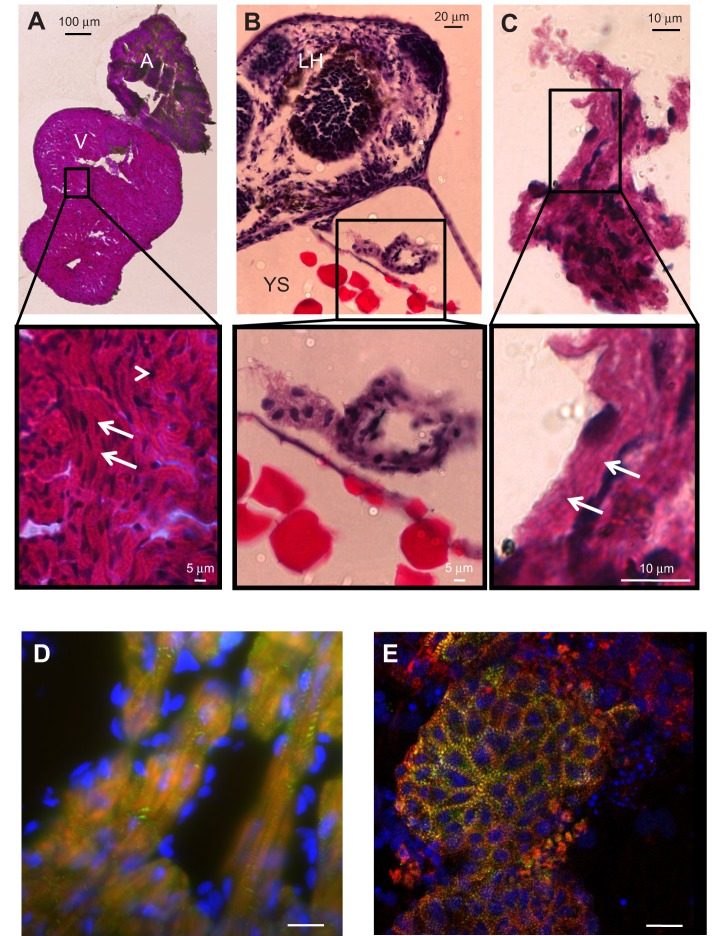
Fig. 2.
Histological and immunohistochemical imaging of ZF adult heart, larval heart and ZFHA. (A) Haematoxylin and Eosin (H&E) staining of atrium and ventricle of an adult ZF (>1 year old). Notice both chambers are composed of spongy myocardium with a small compact layer surrounding the ventricle. The expanded view shows the nuclei (dark stain, arrowhead) and the organisation of the muscle into myocyte bundles with striations visible (arrows). V, ventricle; A, atrium. (B) Section through the anterior of a 3 dpf larval ZF, with the yolk sac (YS) and larval head (LH) clearly visible. The larval heart is shown at higher magnification below. Nuclei are clearly visible, but the muscle structure lacks organisation. (C) ZFHA after 4 days in culture; areas of structural organisation are evident (arrows) and are greater than those of the larval hearts (tissue of origin for ZFHA). (D,E) Immunohistochemical imaging of (D) an adult ZF ventricle and (E) a ZFHA following 3 days in culture stained with α-actinin antibody (green) with actin filaments labelled with phalloidin (red). Nuclei were stained with DAPI (blue). The image in D was captured using epi-fluorescence (Zeiss Axioplan 2 upright microscope), which accounts for the out-of-focus light. The ZFHA image in E has greater resolution as it was captured with a Nikon C1 laser confocal microscope with 488 and 543 nm excitation. In both samples, the α-actinin–actin complex is visible throughout the tissue, highlighting the muscle cell striations. Scale bar, 20 µm for D and E.

