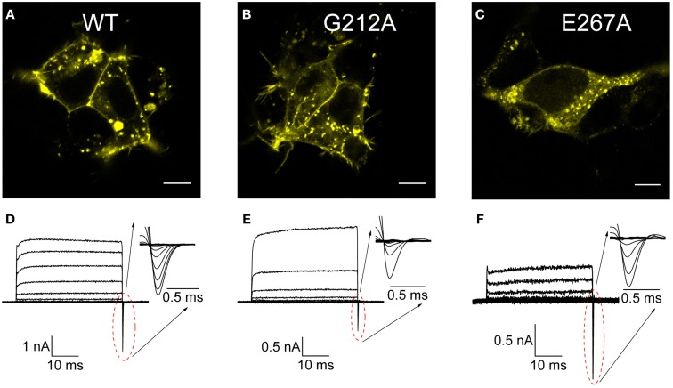
Figure 3.
Cellular localization and transport of WT and disease-associated ClC-5 mutants. (A–C) Confocal florescent images of cells transfected with either WT or the mutants G212A and E267A ClC-5 with fused mCherry at the C-terminus. Visible as bright fluorescent spots is the strong vesicular localization but a significant percentage of the ClC-5 proteins are also localized to the plasma membrane. (D–F) Current families recorded from cells expressing the investigated constructs by whole-cell patch-clamp upon voltage steps between −115 and +175 mV. The insets depict enlarged the off-gating currents for the corresponding mutant that have been used subsequently for estimating the voltage-dependence of ClC-5 activation.

